Motor Learning and Neurological Syndromes Flashcards
(57 cards)
1
Q
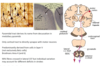
Simple motor pathway
- Upper motorneurone starts in motor cortex of the brain through … ..
- Lower motorneurone starts in the … … cell within spinal cord, moves to NM junction and then …
A
- Upper motorneurone starts in motor cortex of the brain through spinal cord
- Lower motorneurone starts in the anterior horn cell within spinal cord, moves to NM junction and then muscle

2
Q
Upper and Lower motor neuron
A

3
Q
Motor control of tennis serve
A

4
Q
The motor control of hierarchy
A

5
Q
Descending motor pathways
- … - lateral spinal cord
- … - ventro-medial spinal cord
- … tract - carries the volitional control (decision/choice made)
- There are three descending ventromedial pathways as listed.
- These use sensory information about balance, body position and the visual environment to reflexively maintain balance and posture
A
- Voluntary - lateral spinal cord
- Involuntary - ventro-medial spinal cord
- Corticospinal tract - carries the volitional control (decision/choice made)
- There are three descending ventromedial pathways as listed.
- These use sensory information about balance, body position and the visual environment to reflexively maintain balance and posture

6
Q
Descending motor pathways
- Voluntary - lateral spinal cord
- Involuntary - ventro-medial spinal cord
- Corticospinal tract - carries the … control (decision/choice made)
- There are three descending … pathways as listed.
- These use sensory information about balance, body position and the visual environment to reflexively maintain … and ….
A
- Voluntary - lateral spinal cord
- Involuntary - ventro-medial spinal cord
- Corticospinal tract - carries the volitional control (decision/choice made)
- There are three descending ventromedial pathways as listed.
- These use sensory information about balance, body position and the visual environment to reflexively maintain balance and posture

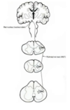
7
Q
The pyramidal or corticospinal tract
- Pyramidal tract derives its name from … in medullary pyramids
- Only cortical tract to directly synapse with … neurons
- Predominantly derived from cells in layer V (not exclusively Betz cells)
- Brodmans Area 4 (and 6)
- …% fibres crossed in lateral CST, …% anterior but individual variation may account for different deficits in strokes
A
- Pyramidal tract derives its name from decussation in medullary pyramids
- Only cortical tract to directly synapse with motor neurons
- Predominantly derived from cells in layer V (not exclusively Betz cells)
- Brodmans Area 4 (and 6)
- 90% fibres crossed in lateral CST, 10% anterior but individual variation may account for different deficits in strokes
8
Q
The pyramidal or corticospinal tract
- Pyramidal tract derives its name from decussation in … pyramids
- Only cortical tract to directly synapse with motor neurons
- Predominantly derived from cells in layer V (not exclusively Betz cells)
- Brodmans Area 4 (and 6)
- 90% fibres crossed in … CST, 10% … but individual variation may account for different deficits in strokes
A
- Pyramidal tract derives its name from decussation in medullary pyramids
- Only cortical tract to directly synapse with motor neurons
- Predominantly derived from cells in layer V (not exclusively Betz cells)
- Brodmans Area 4 (and 6)
- 90% fibres crossed in lateral CST, 10% anterior but individual variation may account for different deficits in strokes

9
Q
Corticospinal tracts
A
- Decussate in medulla - most cross to lateral corticospinal tract before synapse with anterior horn cell

10
Q
Rubrospinal tract
- Unclear to what extend this pathway is involved in humans.
- Predominantly innervates the … muscles in the upper limbs.
- Clinical relevance - low level of … in humans
- … - rubrospinal tract takes over - activated - arm posture change
- Pass in lateral aspect of the spinal cord adjacent to the corticospinal tracts - not voluntary in humans
- Rubrospinal - … nucleus in midbrain - follows tracts - passes through to upper limbs - flexor muscles
A
- Unclear to what extend this pathway is involved in humans.
- Predominantly innervates the flexor muscles in the upper limbs.
- Clinical relevance - low level of arousal in humans
- Stroke - rubrospinal tract takes over - activated - arm posture change
- Pass in lateral aspect of the spinal cord adjacent to the corticospinal tracts - not voluntary in humans
- Rubrospinal - red nucleus in midbrain - follows tracts - passes through to upper limbs - flexor muscles

11
Q
Rubrospinal tract
- Unclear to what extent this pathway is involved in humans.
- Predominantly innervates the flexor muscles in the … limbs.
- Clinical relevance - low level of arousal in humans
- Stroke - rubrospinal tract takes over - activated - … posture change
- Pass in lateral aspect of the spinal cord adjacent to the corticospinal tracts - not voluntary in humans
- Rubrospinal - red nucleus in … - follows tracts - passes through to … limbs - flexor muscles
A
- Unclear to what extend this pathway is involved in humans.
- Predominantly innervates the flexor muscles in the upper limbs.
- Clinical relevance - low level of arousal in humans
- Stroke - rubrospinal tract takes over - activated - arm posture change
- Pass in lateral aspect of the spinal cord adjacent to the corticospinal tracts - not voluntary in humans
- Rubrospinal - red nucleus in midbrain - follows tracts - passes through to upper limbs - flexor muscles
12
Q
Vestibulospinal Tract
- Vestibular organ - 2 aspects
- … - hearing
- … canals - perpendicular to each other - balance
- Medial and lateral tracts
- Originate in the vestibular nuclei of the medulla which relay sensory information from the vestibular labyrinth in the inner ear.
- Medial vestibulospinal pathways projects down to the spinal cord and activates the cervical spinal circuits that control neck and back muscle guides and thus guide … movements.
- Therefore it helps to keeps the … stable as the … is moved.
- Lateral vestibulospinal projects ipsilaterally as far down as the lumbar spinal cord. Helps us maintain an upright and balanced … by facilitating the extensor motor neurons of the legs.
A
- Vestibular organ - 2 aspects
- Cochlear - hearing
- Semicircular canals - perpendicular to each other - balance
- Medial and lateral tracts
- Originate in the vestibular nuclei of the medulla which relay sensory information from the vestibular labyrinth in the inner ear.
- Medial vestibulospinal pathways projects down to the spinal cord and activates the cervical spinal circuits that control neck and back muscle guides and thus guide head movements.
- Therefore it helps to keeps the eyes stable as the body is moved.
- Lateral vestibulospinal projects ipsilaterally as far down as the lumbar spinal cord. Helps us maintain an upright and balanced posture by facilitating the extensor motor neurons of the legs.

13
Q
Vestibulospinal Tract
- Vestibular organ - 2 aspects
- Cochlear - hearing
- Semicircular canals - perpendicular to each other - balance
- Medial and lateral tracts
- Originate in the vestibular nuclei of the medulla which relay sensory information from the vestibular labyrinth in the inner ear.
- … vestibulospinal pathways projects down to the spinal cord and activates the cervical spinal circuits that control neck and back muscle guides and thus guide head movements.
- Therefore it helps to keeps the eyes stable as the body is moved.
- … vestibulospinal projects ipsilaterally as far down as the lumbar spinal cord. Helps us maintain an upright and balanced posture by facilitating the extensor motor neurons of the legs.
A
- Vestibular organ - 2 aspects
- Cochlear - hearing
- Semicircular canals - perpendicular to each other - balance
- Medial and lateral tracts
- Originate in the vestibular nuclei of the medulla which relay sensory information from the vestibular labyrinth in the inner ear.
- Medial vestibulospinal pathways projects down to the spinal cord and activates the cervical spinal circuits that control neck and back muscle guides and thus guide head movements.
- Therefore it helps to keeps the eyes stable as the body is moved.
- Lateral vestibulospinal projects ipsilaterally as far down as the lumbar spinal cord. Helps us maintain an upright and balanced posture by facilitating the extensor motor neurons of the legs.

14
Q
Tectospinal Tract
- Originates in the superior … in the midbrain which receives direct input from the …
- The superior … receives information from the … and the visual cortex. This is used to construct a map of the world around us.
- Allows us to direct the head and eyes to … so that the appropriate point of space is imaged on the …
- The projections decussate immediately and lie close to the midline into the cervical regions of the spinal cord where they help to control the muscles of the neck, upper trunk and shoulders.
A
- Originates in the superior colliculus in the midbrain which receives direct input from the retina.
- The superior colliculus receives information from the retina and the visual cortex. This is used to construct a map of the world around us.
- Allows us to direct the head and eyes to move so that the appropriate point of space is imaged on the fovea.
- The projections decussate immediately and lie close to the midline into the cervical regions of the spinal cord where they help to control the muscles of the neck, upper trunk and shoulders.

15
Q
Tectospinal Tract
- Originates in the … … in the midbrain which receives direct input from the retina.
- The … … receives information from the retina and the visual cortex. This is used to construct a map of the world around us.
- Allows us to direct the head and eyes to move so that the appropriate point of space is imaged on the fovea.
- The projections … immediately and lie close to the midline into the cervical regions of the spinal cord where they help to control the muscles of the neck, upper trunk and shoulders.
A
- Originates in the superior colliculus in the midbrain which receives direct input from the retina.
- The superior colliculus receives information from the retina and the visual cortex. This is used to construct a map of the world around us.
- Allows us to direct the head and eyes to move so that the appropriate point of space is imaged on the fovea.
- The projections decussate immediately and lie close to the midline into the cervical regions of the spinal cord where they help to control the muscles of the neck, upper trunk and shoulders.

16
Q
Reticulospinal Tract
- … - arousal state - 2 tracts, medial and lateral -> … muscles -> balance
- The pathway runs from the … The reticular formation is just under the cerebral aqueduct and fourth ventricle. It is a complex meshwork of neurons.
- It descends in two separate pathways, pontine (medial) and medullary (lateral) .
- Both facilitate the … of the limbs.
A
- Involuntary - arousal state - 2 tracts, medial and lateral -> extensor muscles -> balance
- The pathway runs from the brainstem. The reticular formation is just under the cerebral aqueduct and fourth ventricle. It is a complex meshwork of neurons.
- It descends in two separate pathways, pontine (medial) and medullary (lateral) .
- Both facilitate the extension of the limbs.

17
Q
Reticulospinal Tract
- Involuntary - arousal state - 2 tracts, … and… -> extensor muscles -> balance
- The pathway runs from the brainstem. The reticular formation is just under the cerebral aqueduct and fourth ventricle. It is a complex meshwork of neurons.
- It descends in two separate pathways, pontine (…) and medullary (…) .
- Both facilitate the extension of the limbs.
A
- Involuntary - arousal state - 2 tracts, medial and lateral -> extensor muscles -> balance
- The pathway runs from the brainstem. The reticular formation is just under the cerebral aqueduct and fourth ventricle. It is a complex meshwork of neurons.
- It descends in two separate pathways, pontine (medial) and medullary (lateral) .
- Both facilitate the extension of the limbs.

18
Q
Overview of descending pathways
- Tectospinal and medial vestibulospinal
- Control … and … movements.
- Lateral vestibulospinal and reticulospinal
- Activate … muscles in arms and legs.
- Rubrospinal
- Activates … muscles in arms.
A
- Tectospinal and medial vestibulospinal
- Control head and neck movements.
- Lateral vestibulospinal and reticulospinal
- Activate extensor muscles in arms and legs.
- Rubrospinal
- Activates flexor muscles in arms.
19
Q
Overview of descending pathways
- T… and … vestibulospinal
- Control head and neck movements.
- Lateral … and …
- Activate extensor muscles in arms and legs.
- R…
- Activates flexor muscles in arms.
A
- Tectospinal and medial vestibulospinal
- Control head and neck movements.
- Lateral vestibulospinal and reticulospinal
- Activate extensor muscles in arms and legs.
- Rubrospinal
- Activates flexor muscles in arms.
20
Q
OVERVIEW OF THE DESCENDING PATHWAYS
A

21
Q
Descending tracts - posturing in coma
- … coma scale
A
- Glasgow coma scale
22
Q
Glasgow coma scale
- Minimum of … - … max
- Motor response - noxious stimuli if no response to talking - trigger - response in limbs - tell from response to … where lesion is
- … posturing: 2 points - Above red nucleus - decorticate posturing - stroke - corticospinal tract damaged but rubrospinal tract intact - activate flexor muscle
- … posturing: 1 point - Below red nucleus - inferior to red nucleus - lose rubrospinal tracts - lateral vestibular and reticulospinal tract - involved in extensors - arm and legs - all 4 legs - worse prognosis below red nucleus

A
- Glasgow coma scale
- Minimum of 3 - 15 max
- Motor response - noxious stimuli if no response to talking - trigger - response in limbs - tell from response to pain where lesion is
- Decorticate posturing: 2 points - Above red nucleus - decorticate posturing - stroke - corticospinal tract damaged but rubrospinal tract intact - activate flexor muscle
- Decerebrate posturing: 1 point - Below red nucleus - inferior to red nucleus - lose rubrospinal tracts - lateral vestibular and reticulospinal tract - involved in extensors - arm and legs - all 4 legs - worse prognosis below red nucleus
23
Q
Glasgow coma scale
- Minimum of 3 - 15 max
- Motor response - noxious stimuli if no response to talking - trigger - response in limbs - tell from response to pain where lesion is
- Decorticate posturing: … points - … red nucleus - decorticate posturing - stroke - corticospinal tract damaged but rubrospinal tract intact - activate flexor muscle
- Decerebrate posturing: … point - … red nucleus - inferior to red nucleus - lose rubrospinal tracts - lateral vestibular and reticulospinal tract - involved in extensors - arm and legs - all 4 legs - worse prognosis … red nucleus

A
- Glasgow coma scale
- Minimum of 3 - 15 max
- Motor response - noxious stimuli if no response to talking - trigger - response in limbs - tell from response to pain where lesion is
- Decorticate posturing: 2 points - Above red nucleus - decorticate posturing - stroke - corticospinal tract damaged but rubrospinal tract intact - activate flexor muscle
- Decerebrate posturing: 1 point - Below red nucleus - inferior to red nucleus - lose rubrospinal tracts - lateral vestibular and reticulospinal tract - involved in extensors - arm and legs - all 4 legs - worse prognosis below red nucleus
24
Q
Glasgow coma scale
- Motor response:
- Rubrospinal pathways are the key:
- In decorticate posturing, the rubrospinal are disinhibited and therefore facilitate flexors in the UL (lesion … red nucleus)
- In decerebrate posturing, the rubrospinal are disrupted and therefore the UL are extended. (lesion … red nucleus)
- Red nucleus is in the …
- So response to noxious stimulus allows us to understand where the lesion is.
- Lateral vestibulospinal and reticulospinal tracts activate the … muscles.
A
- Rubrospinal pathways are the key:
- In decorticate posturing, the rubrospinal are disinhibited and therefore facilitate flexors in the UL (lesion above red nucleus)
- In decerebrate posturing, the rubrospinal are disrupted and therefore the UL are extended. (lesion below red nucleus)
- Red nucleus is in the midbrain
- So response to noxious stimulus allows us to understand where the lesion is.
- Lateral vestibulospinal and reticulospinal tracts activate the extensor muscles.
25
_Damage to motor cortex and corticospinal tract - humans_
* During ... - damage corticospinal tract
* Extensor muscle from other tracts - reticulospinal and vestibulospinal cause extensors to lower limb - Can still walk
* ... neurone problem can be picked up against lower neurone problem
* Upper - taken brakes off, reflexes more brisk
* Increased tone in arms and increased reflexes in upper motor neurone problem
* Typical Posture
* some preserved upper limb flexion
* and lower limb extension
* ... tone (spasticity), ... Reflexes,
* Extensor Plantar/Babinski reflex, Clonus
* But patient maintains a posture
* During **stroke** - damage corticospinal tract
* Extensor muscle from other tracts - reticulospinal and vestibulospinal cause extensors to lower limb - Can still walk
* **Upper** neurone problem can be picked up against lower neurone problem
* Upper - taken brakes off, reflexes more brisk
* Increased tone in arms and increased reflexes in upper motor neurone problem
* Typical Posture
* some preserved upper limb flexion
* and lower limb extension
* **Increased tone (spasticity), Brisk Reflexes,**
* Extensor Plantar/Babinski reflex, Clonus
* But patient maintains a posture

26
_Damage to motor cortex and corticospinal tract - humans_
* During stroke - damage corticospinal tract
* Extensor muscle from other tracts - reticulospinal and vestibulospinal cause extensors to lower limb - Can still walk
* Upper neurone problem can be picked up against lower neurone problem
* Upper - taken brakes off, reflexes more brisk
* Increased tone in arms and increased reflexes in upper motor neurone problem
* Typical Posture
* some preserved upper limb flexion
* and lower limb extension
* Increased tone (spasticity), Brisk Reflexes,
* Extensor Plantar/Babinski reflex, Clonus
* But patient maintains a posture

27
Increased tone in arms and increased reflexes in ... motor neurone problem
Increased tone in arms and increased reflexes in **upper** motor neurone problem
28
_Loss of descending inhibition_
* Altered excitability of spinal inhibitory interneurons
* ... reflexes
* ... tone to rapid passive muscle stretching= spasticity
* Contrast with damage to motor neuron
* – reduced tone
* ... of reflexes
* muscle ...
* Babinksi reflex - A-normal (Flexor) B-abnormal (extensor)
* Altered excitability of spinal inhibitory interneurons
* Brisk reflexes
* increased tone to rapid passive muscle stretching= spasticity
* Contrast with damage to motor neuron
* – reduced tone
* loss of reflexes
* muscle wasting
* Babinksi reflex - A-normal (Flexor) B-abnormal (extensor)

29
_Loss of descending inhibition_
* Altered excitability of spinal inhibitory interneurons
* Brisk reflexes
* increased tone to rapid passive muscle stretching= ...
* Contrast with damage to motor neuron
* – ... tone
* loss of reflexes
* muscle wasting
* ... reflex - A-normal (Flexor) B-abnormal (extensor)
* Altered excitability of spinal inhibitory interneurons
* Brisk reflexes
* increased tone to rapid passive muscle stretching= spasticity
* Contrast with damage to motor neuron
* – reduced tone
* loss of reflexes
* muscle wasting
* Babinksi reflex - A-normal (Flexor) B-abnormal (extensor)

30
When differentiating upper and lower motor neuron disease, remember that ... motor neurons are responsible for motor movement, whereas ... motor neurons prevent excessive muscle movement. ... motor disorders usually cause spasticity; ... motordisorders usually cause flaccidity.
When differentiating upper and lower motor neuron disease, remember that upper motor neurons are responsible for motor movement, whereas lower motor neurons prevent excessive muscle movement. Upper motor disorders usually cause spasticity; lower motordisorders usually cause flaccidity.
31
_Babinski Reflex_
* In a baby, toes ... and ...
* As corticospinal tract develops - tickle, toes go ...
* ... motor neurone problem - reverts back into what you see in a baby
* Babinski ... - shows you have a problem
* In a baby, toes lift and fan
* As corticospinal tract develops - tickle, toes go down
* Upper motor neurone problem - reverts back into what you see in a baby
* Babinski positive - shows you have a problem

32
Babinski + = ... motor neuron problem
Babinski + = **upper** motor neuron problem

33
_Corticobulbar pathway_
* The ... tract carries motor signals from the primary motor cortex in the brain, down the spinal cord, to the muscles of the trunk and limbs.
* The ... tract carries efferent, motor, information from the primary motor cortex to the muscles of the face, head and neck.
* Axons also project from layer 5 to the motor neurons in the brainstem.
* The **corticospinal** tract carries motor signals from the primary motor cortex in the brain, down the spinal cord, to the muscles of the trunk and limbs. ...
* The **corticobulbar** tract carries efferent, motor, information from the primary motor cortex to the muscles of the face, head and neck.
* Axons also project from layer 5 to the motor neurons in the brainstem.

34
The ... tract is a descending pathway responsible for innervating several cranial nerves, and runs in paralell with the corticospinal tract.
The **corticobulbar** tract is a descending pathway responsible for innervating several cranial nerves, and runs in paralell with the corticospinal tract.
35
_Stroke - facial palsy_
* Problem with left hemisphere - right side weakness
* Decussation of the cortical spinal tracts
* For the face - stroke - ... part of the face is bilaterally innervated
* Lose one side, ... face is spared
* Only see weakness of contralateral lower side of the face
* Distinguish from disorder of lower motor nerve of cranial nerve 7 - facial nerve - take out whole side of the face
* Stroke mimics - ... palsy - eyes and mouth weakness
* Stroke - only mouth - eyes are sparred
* Problem with left hemisphere - right side weakness
* Decussation of the cortical spinal tracts
* For the face - stroke - upper part of the face is bilaterally innervated
* Lose one side, upper face is spared
* Only see weakness of contralateral lower side of the face
* Distinguish from disorder of lower motor nerve of cranial nerve 7 - facial nerve - take out whole side of the face
* Stroke mimics - bell's palsy - eyes and mouth weakness
* Stroke - only mouth - eyes are sparred

36
_Stroke - facial palsy_
* Problem with left hemisphere - right side weakness
* Decussation of the cortical spinal tracts
* For the face - stroke - upper part of the face is bilaterally innervated
* Lose one side, upper face is spared
* Only see weakness of contralateral lower side of the face
* Distinguish from disorder of lower motor nerve of cranial nerve 7 - facial nerve - take out whole side of the face
* Stroke mimics - bell's ... - eyes and mouth weakness
* Stroke - only mouth - eyes are ...
* Problem with left hemisphere - right side weakness
* Decussation of the cortical spinal tracts
* For the face - stroke - upper part of the face is bilaterally innervated
* Lose one side, upper face is spared
* Only see weakness of contralateral lower side of the face
* Distinguish from disorder of lower motor nerve of cranial nerve 7 - facial nerve - take out whole side of the face
* Stroke mimics - bell's palsy - eyes and mouth weakness
* Stroke - only mouth - eyes are sparred

37
The motor ... is a topographic representation of the body parts and its correspondents along the precentral gyrus of the frontal lobe. While the sensory ... is a topographic representation of the body parts along the postcentral gyrus of the parietal lobe.
The motor homunculus is a topographic representation of the body parts and its correspondents along the precentral gyrus of the frontal lobe. While the sensory homunculus is a topographic representation of the body parts along the postcentral gyrus of the parietal lobe.
38
This is showing a ...sagittal meningioma - by pressing of foot/leg area of both motor cortices it presents as ... leg weakness and spasticity

This is showing a parasagittal meningioma - by pressing of foot/leg area of both motor cortices it presents as bilateral leg weakness and spasticity
39
_Blood supply to the brain and stroke syndromes_
* Always deficits follow vascular territory
* Revise blood supply to hemisphere - label


40
_Middle Cerebral artery occlusion_
* MCA usually hand and face but depends where it is
* Proximal lesion - corticospinal tract starts in cortex but travels down - merge in ... capsule - take out leg, hand, face
* Can get with MCA - if its deep - undercutting as it travels through internal capsule
* ... lesion - only face and hand, maybe language function
* Learn ... - nerves coming to ... capsule
* MCA usually hand and face but depends where it is
* Proximal lesion - corticospinal tract starts in cortex but travels down - merge in internal capsule - take out leg, hand, face
* Can get with MCA - if its deep - undercutting as it travels through internal capsule
* Distal lesion - only face and hand, maybe language function
* Learn homunculus - nerves coming to internal capsule

41
_Anterior cerebral artery stroke_
* Supplies medial part of ... lobes including leg area of motor cortex
* crural (leg) paresis \> arm paresis
* frontal signs (A.g. ... - loss of impairment of the ability to make decisions or act independently)
* Supplies medial part of frontal lobes including leg area of motor cortex
* crural (leg) paresis \> arm paresis
* frontal signs (A.g. abulia - loss of impairment of the ability to make decisions or act independently)

42
_Anterior cerebral artery stroke_
* Supplies medial part of frontal lobes including ... area of motor cortex
* crural (leg) paresis \> arm paresis
* frontal signs (A.g. abulia - loss of ... of the ability to make ... or act ...)
* Supplies medial part of frontal lobes including leg area of motor cortex
* crural (leg) paresis \> arm paresis
* frontal signs (A.g. abulia - loss of impairment of the ability to make decisions or act independently)

43
_Jacksonian seizure_
* Pure ... seizure - start in one part - spread up arm and involve face
* Partial onset simple ... seizure becoming secondarily generalised
* Strongly associated with structural abnormality in or close to ... cortex
* Pure motor seizure - start in one part - spread up arm and involve face
* Partial onset simple motor seizure becoming secondarily generalised
* Strongly associated with structural abnormality in or close to motor cortex

44
_Posterior Parietal Cortex_
* Posterior parietal cortex - area ... and ... - Info around you - need sensory feedback while walking etc
* Sensory ... - problems - unsteady - problem with sensory nerves - must have sensory feedback to have good ...
* Feeds to sensory cortex
* Posterior parietal cortex - area 5 and 7 - Info around you - need sensory feedback while walking etc
* Sensory neuropathy - problems - unsteady - problem with sensory nerves - must have sensory feedback to have good balance
* Feeds to sensory cortex

45
Various sensations - visual, a..., h..., touch, vestibular ..., ... taste
**Various sensations - visual, auditory, hearing, touch, vestibular balance, gustatory taste**
46
_Perceptual motor dysfunction_

47
_Association cortices_
* ... - sensation
* ... - vision
* ... - vestibular and hearing
* All feeds to frontal lobe
* Frontal lobe - p...
* ... cortex - SMA and PMA
* Parietal - sensation
* Occipital - vision
* Temporal - vestibular and hearing
* All feeds to frontal lobe
* Frontal lobe - planning
* Prefrontal cortex - SMA and PMA

48
_Premotor area (PMA)_
* Importance in control of ... ... movements - e.g. orientation of hand in relation to object to be ... (prehension)
* Damage may also cause perseveration of motor activity despite lack of success
* Importance in control of visually guided movements - e.g. orientation of hand in relation to object to be grasped (prehension)
* Damage may also cause perseveration of motor activity despite lack of success

49
_Simple finger flexion - only M1 activation_
* Functional imaging - highlights activated brain area
* Flex finger - M1 activated
* Then a sequence of movements - M1+ SMA activated - more complex movement
* But if mental ... only of finger movements - only SMA activation
* ... takes place in SMA

* Functional imaging - highlights activated brain area
* Flex finger - M1 activated
* Then a sequence of movements - M1+ SMA activated - more complex movement
* But if mental rehearsal only of finger movements - only SMA activation
* Thought takes place in SMA
50
In humands - wide interconnections between sensory and motor association areas - damage causes ...
In humands - wide interconnections between sensory and motor association areas - damage causes apraxia
51
In humands - wide interconnections between sensory and motor association areas - damage causes apraxia - this means the inability to carry out ... movements in the absence of ... or ...
In humands - wide interconnections between sensory and motor association areas - damage causes apraxia - this means the inability to carry out purposeful movements in the absence of paralysis or paresis
52
_Types of Apraxia_
* ... (parietal): unable to report the sequence - unable to ... how to do it
* Show me how to make a peanut butter sandwich?
* ... (SMA): unable to use the tool - unable to actually ... the tool
* Show me how to hold and use a pair of scissors
* Ideational (parietal): unable to report the sequence - unable to explain how to do it
* Show me how to make a peanut butter sandwich?
* Ideomotor (SMA): unable to use the tool - unable to actually use the tool
* Show me how to hold and use a pair of scissors
53
_Types of Apraxia_
* Ideational (...): unable to report the sequence - unable to explain how to do it
* Show me how to make a peanut butter sandwich?
* Ideomotor (...): unable to use the tool - unable to actually use the tool
* Show me how to hold and use a pair of scissors
* Ideational (parietal): unable to report the sequence - unable to explain how to do it
* Show me how to make a peanut butter sandwich?
* Ideomotor (SMA): unable to use the tool - unable to actually use the tool
* Show me how to hold and use a pair of scissors
54
_Aberrant sensory processing - task specific dystonias_

55
_Role of anterior cingulate gyrus_
* Pathway from anterior cingulate gyrus - can ... for a genuine joke
* Even during a ...
* Pathway from anterior cingulate gyrus - can smile for a genuine joke
* Even during a stroke
56
_Basal Ganglia_
* Function: ... feedback loop with the cortex to select ... movements and deselect ... movements.
* Function: Positive feedback loop with the cortex to select wanted movements and deselect unwanted movements.

57
_Cerebellum_
* Function:
* ... of muscles in order to make smooth movements.
* B...
* ... learning
* Function:
* Coordination of muscles in order to make smooth movements.
* Balance
* Motor learning

