Basal Ganglia Flashcards

(47 cards)

1
Q

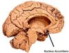
Location of the nucleus accumbens

A

In the most rostral and ventral part of the corpus striatum

2
Q

Corpus striatum

A

=basal ganglia

basal nuclei

Caudate

Putamen

Globus pallidus

3
Q

Relationship between the basal ganglia and the amygdala

A

Similar embryological derivation but functionally different.

The nucleus accumbens also has close connections with the amygdala, thus providing an important link between the BG and the limbic system

4
Q
A
5
Q
A
6
Q

Nucleus accumbens function

A

Associated with reward and gratification.

It is an important site of action for addictive substances

7
Q

Lentiform nucleus=

A

Putamen and GP

Point lies against the genu of the internal capsule.

8
Q

With what is the putamen most closely associated on phylogenetic, connectional and functional grounds?

A

Caudate nucleus rather than GP

9
Q

Paleostriatum

A

=GP

10
Q

Neostriatum=

A

Caudate and putamen

11
Q

Striatum=

A

Putamen and caudate nucleus

12
Q
A
13
Q

What separates the putamen from the globus pallidus?

A

A thin lamina of nerve fibres- the lateral medullary lamina

14
Q

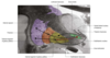
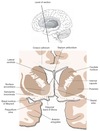
Layers of white matter and grey matter from insula to caudate

A

Insula

Extreme capsule

Clasutra

External capsule

Putamen

Lateral medullary lamina

GP

15
Q

Relationship of the caudate to the putamen

A

At the head of caudate it is almost completely separated from the putamen by the internal capsule

At its rostral extermity, it is continuous with the putamen through and beneath the anterior limb of the internal capsule.

At this anterior level, the most ventral and medial part of the striatum constitutes the nucleus accumbens.

16
Q

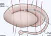
Relationship of the caudate nucleus to the ventricular system

A

Head of the caudate causes a prominent bulge in the lateral wall of the anterior horn of the lateral ventricle.

It also passes posteriorly, gradually tapering and following the curvature of the ventricle, descending into the temporal lobe where it lies in the roof of the inferior horn

17
Q

Anatomical arrangement of the globus pallidus

A

External

Medial medullary lamina

Internal

18
Q

Relationship of the GPi

A

Shares many similarities in cytology and connections with the pars reticulata of the SN in the midbrain.

Although the two are separated by the internal capsule, these are best regarded as a single entity in the functional sense.

19
Q

Substantita innominata

A

Refers to the basal part of the rostral forebrain that lies beneath the corpus stritatum.

This complex region contains several groups of neurones, one of them being the nucleus basalis of meynert that project widely to the cerebral cortex and utilise ACh as their neurotransmitter.

These neurones undergo degeneration in Alzheimer’s

20
Q

What are the three basal ganglia loops of the striatum

A

Putamen- voluntary motor

Caudate- volunatry eye movements, cognitive-executive

Ventral striatum- emotional affective

21
Q

Input portion of the basal ganglia

22
Q

What are the three principle sources of striatal afferents?

A

Cerebral cortex

Thalamus

SN

23
Q

Corticostriatal fibres

A

Origin from widespread regions of the cerebral cortex, predominantly on the ipsilateral lobe.

Motor project mainly to the putamen

More anterior regions of the frontal lobe and other association cortices project mainly to the caudate.

Corticostriatal fibres are excitatory and use glutamic acid as their neurotransmitter

24
Q

Thalamostriatal projection

A

Come from intralaminar nuclei (centromedian and parafascicular nuclei) of the ipsilateral thalamus

25
Nigrostriatal projection
From the pars compacta of the ipsilateral SN of the midbrain tegmentum Dopaminergic which has both excitatory and inhibitory effects.
26
Pars compacta
Portion of the SN which contains the dark pigment neuromelanin which is a by-product of dopamine synthesis
27
Whence does the nucleus accumbens receive its afferents?
From the ventral tegmental area, which lies medial to the SN. This projection is part of the mesolimbic pathway which also provides dopaminergic innervation of the amygdala.
28
Efferent projections of the striatum
Directed principally to the two segments of the globus pallidus and pars reticulata of the SN (striatopallidal and striagonigral fibres)
29
Striatal efferent output
Inhibitory upon pallidal and nigral neurones with GABAergic neurotransmission. A number of neuropeptides are colocalised in these efferent neurones. The cells that porject to the internal segment of the globus pallidus and SN contain substance P and dynoprhin. The projection to the external segment of the GP contains met-enkephalin
30
Ouput portion of the basal ganglia?
The internal pallidum and pars reticulata of the substantia nigra as they are the origin of the majority of basal ganglia efferent fibres
31
Pallidal afferents
Arise principally from the striatum and subthalamic nucleus. Both utilise GABA as thier primary transmitter. Coefferents projecting to GPe contain enkephalin while those projecting to GPi contain substance P and dynorphin
32
Subthalamopallidal projection
Originates in the STN of the caudal diencephalon Located beneath the thalamus against the medial border of the internal capsule. These fibres pass laterally through the internal capsule contributing to the subthalamic fasciculus, terminating in both segments of the globus pallidus
33
34
35
36
37
38
Output of subthalamopallidal pathway
Excitatory to pallidal neurones, using glutamic acid as its transmitter. The STN also sends similar fibres to the pars reticulata of the substantia nigra.
39
GPe efferents
Principally on the STN Pallidosubthalamic fibres pass medially through the internal capsule to the subthalamic fasciculus. This projection is inhibitory, using GABA as its transmitter
40
GPi efferents
Along with the pars reticulata of the SN projects primarily to the thalamus with a smaller projection to the brainstem tegmentum. These output neurones are all inhibitory and utilise GABA as their transmitter
41
What are the two possible routes of palladothalamic fibres?
Ansa lenticularis (around the anterior marigin of the internal capsule) Lenticular fasciulus (through the internal capsule). These fibres course medially and then loop dorsally and latearlly as the thalamic fasciculus. In following this trajectory, the pallidothalamic fibres circumnavigate a cellular region of the subthalamus known as the zona incerta, which lies between the thalamus and the STN
42
What are the main targets of pallidothalamic fibres?
Ventral anterior and ventral lateral thaalamic nuclei which project glutamatergic fibres to the motor regions of the frontal lobe A smaller contingent of medial pallidal efferent fibres pass caudally to termiante in the brainstem tegmentum in the nucleus tegmenti pedunculopontinus which lies at the boundary between the midbrain and the pons, near the lateral edge of the superior cerebellar peduncle. This area has been termed the mesencephalic locomotor region.
43
Somatotropic organisaiton of striatal projections to the GP and SN
Pallidal neuroens are primarily involved in limb movements. Nigral cells control the axial muscualture including the EOM.
44
Role of BG in movement
Cortical efferents to striatum causing ecxitation. Direct pathway- directly inihbiting internal pallidal or pars reticulata neurones. Causing disinhibition of target neurones and subsequent excitation of motor cortical neuroens. Indirect pathway- via the STN. Efferents terminate int he GPe inducing inihibtion of GPe inhibitory neurones disinhibiting the subthalamic neuroens. The resultant increase in discharge of STN causes activation of internal pallidal and nigral neuroens inihibting thalamic and cortical cells. Inhibiting unwanted movmenents.
45
Draw the basal ganglia motor control pathway
46
Action of dopamine on BG
Exerts an exciatory influence on the direct pathway i.e. disinihbiting movement. Inhibitory effect on indirect pathway.
47
The ‘direct’ striatal output pathway: Projects to the external globus pallidus (gpe) Uses glutamate as its neurotransmitter Contains the neuropeptide enkephalin Inhibits the subthalamic nucleus Originates from ‘medium spiny’ striatal neurones
The ‘direct’ striatal output pathway originates from medium spiny neurones and projects to the internal globus pallidus (GPi). It uses GABA as its neurotransmitter and also contains dynorphin/substance P.