Questions Flashcards
(21 cards)
The spinal nucleus of the trigeminal nerve is continuous with which of the following structures?
Select one:
a. The accessory cuneate nucleus
b. The spinothalamic tract
c. The substantia gelatinosa
d. The lateral lemniscus
e. The ventral cochlear nucleus
The spinal nucleus of the trigeminal nerve receives pain and temperature input from the face. This is continuous with the substantia gelatinosa in the spinal cord, which is the first point of synapse for pain fibres in the spinal cord. The lateral lemniscus is formed by projections of the ventral cochlear nucleus on the way to the inferior collicullus.
The correct answer is: The substantia gelatinosa
Which of the following is the best description of the anterior commisure?
Select one:
a. Connects both temporal lobes
b. It forms the inferior boundary of the pineal recess
c. It forms the posterior part of the lamina terminalis
d. It is found in the lateral walls of the third ventricle
e. It is found in the rostrum of the corpus callosum
The anterior commissure is connects both temporal lobes. It forms the superior border of the lamina terminalis on the anterior wall of the third ventricle.
The correct answer is: Connects both temporal lobes
The internal olivary complex receives information regarding proprioception and fine touch discrimination via which structure?
Select one:
a. Dentate nucleus
b. Inferior cerebellar peduncle
c. External arcuate fibres
d. Internal arcuate fibres
e. Lateral lemniscus
The internal olivary complex receives proprioceptive input via internal arcuate fibres. These inputs are then relayed as climbing fibres to the cerebellar cortex. The internal arcuate fibres also form ventral projections from the gracillis and cuneate nuclei that form the medial lemniscus in the medulla. The external arcuate fibres form the cuneocerebellar tract to ascend the inferior cerebellar peduncle without entering the olivary complex.
The correct answer is: Internal arcuate fibres
7-year-old boy is admitted following with reduced consciousness and fever. He receives a head CT demonstrating cerebral oedema. Which finding would be consistent with cerebral oedema?
Select one:
a. Patent basal cisterns
b. Hyperattenuated cerebellum
c. Hyperattenuation of the white matter
d. Patent third ventricle
e. Enlarged temporal horns
Cerebral oedema is associated with a hyperdense cerebellum in contrast to a hypodense forebrain. Enlarged temporal horns and patent third ventricles may be seen in hydrocephalus. Cerebral oedema may result in encroachment upon the basal cisterns, which may leads to concerns regarding the safety of the brain stem.
The correct answer is: Hyperattenuated cerebellum
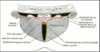
A 2-year-old child is reffered for 3rd ventriclostomy. Which of the following structures can be visualized on the floor of the third ventricle using a transcortical endoscopic approach.
Select one:
a. Septal vein
b. The body of the fornix
c. Pituitary stalk
d. Optic cistern
e. Area vasculosa
The view of the floor of the third ventricle from an endoscope demonstrates the area vasculosa anteriorly and the mammilllary bodies posteriorly. The body of the fornix forms the roof of the third ventricle. The pituitary stalk is not visualized within the 3rd venticle. The septal vein is visualized in the lateral ventricle of during endoscopic 3rd ventriclostomy.
The correct answer is: Area vasculosa
65-year-old patient with a past medical history of atrial fibrillation is brought to the emergency department following collapse. On arrival, his eyes deviate to the right and he has a left-sided spastic hemiplegia affecting both his left arm and leg. 3-days post-admission the patient died after developing increased intracranial pressure resulting in brain herniation. What kind of herniation is likely in this patient?
Select one:
a. Subfalcine herniation
b. Transtentorial herniation
c. Upward herniation
d. Tonsillar herniation
e. Central herniation
This patient has likely suffered a stroke due to occlusion of the left middle cerebral artery. Cerebral oedema is common sequela of ischaemia. In middle cerebral artery stroke oedematous cerebral hemisphere can herniate across the falx cerbri.
The correct answer is: Subfalcine herniation
Which venous system lies within the tela chorioidea?
Select one:
a. Vein of Trolard
b. Internal cerebral veins
c. The great cerebral vein
d. Thalamostriate veins
e. The veins of Rosenthal
The great cerebral vein is formed by the internal cerebral veins and the veins of Rosenthal. The tela chorioidea is a double layer of pia between the fornix, above, and the cavity of the third ventricle, below. Within the tela chorioidea are the internal cerebral veins.
The correct answer is: Internal cerebral veins
A 36-year-old man presents with sudden onset headache. A head CT reveals hyperdensity in the basal cisterns and the lateral ventricles. Cerebral angiography reveals a ruptured anterior communicating artery aneurysm. What explains the presence of blood in the lateral ventricles in this patient?
Select one:
a. Periventricular heamorrhage
b. Bleeding of the choroid plexus
c. Rupturing through the tuber cinerium
d. Retrograde flow through the foramen of sylvius
e. Rupturing through the lamina terminalis
In subarachnoid haemorrhage, blood enters the lateral ventricles by rupturing through the lamina terminalis.
The correct answer is: Rupturing through the lamina terminalis
Hyposmia, hypogonadism and short stature is a disorder secondary to migration failure of hypothalamic neurones. With what syndrome is this disorder associated?
Select one:
a. Wallenburg’s syndrome
b. Foster-Kennedy Syndrome
c. Kallman’s syndrome
d. Benedikt’s syndrome
e. Neurofibromatosis
Kallman’s Syndrome is due to migration failure of hypothalamic neurons resulting in reduced pituitary function and an absent olfactory bulb. Foster-Kennedy syndrome is caused by a anterior fossa skull base meningioma causing anosmia, ipsilateral optic atrophy and contralateral papilloedema.
The correct answer is: Kallman’s syndrome
The amygdala receives input from the olfactory nerve via which of the following structures?
Select one:
a. The anterior perforating substance
b. The thalamus
c. The medial olfactory stria
d. The lateral olfactory stria
e. The olfactory trigone
The amygdala receives input from the lateral olfactory stria. Olfaction is the only sensation that does not relay through the thalamus. The olfactory nerve divides into medial and lateral stria forming a trigone, within that triangle is the anterior perforating substance.
The correct answer is: The lateral olfactory stria
A patient develops left sided facial droop. The patient also has a vesicular rash in the external auditory meatus and the hard palate. The patient is diagnosed with Ramsey Hunt Syndrome. What best described the dormant site of the Varicella Zoster Virus in this patient?
Select one:
a. The dorsal root ganglion of C2
b. The otic ganglion
c. The pterygopalatine gangion
d. The facial nucleus
e. The geniculate ganglion
Varicella Zoster lies dormant in the dorsal root ganglia, which can reactivate causing a unilateral vesicular rash along the distribution of its respective nerve. In Ramsey Hunt syndrome the facial nerve is affected where the virus lies dormant in the geniculate ganglion. The geniculate, knee–‐shaped, ganglion lies within the facial canal and giving rise to the lesser and greater petrosal nerves and the facial nerve.
The correct answer is: The geniculate ganglion
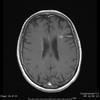
A 34-year-old woman is brought to A&E. She delivered her first baby 7 days ago. The neurosurgical registrar has noted the presence of empty delta sign on her head CT. What finding is consistent with empty delta sign?
Select one:
a. Bilateral glioma
b. Superior sagittal sinus thrombosis
c. Periventricular perivenular inflammation
d. Intraparenchymal venous abnormality with radiating veins
e. Acute clot formation within the middle cerebral artery
A-E describe eponymous terms in neuroradiology. This empty delta sign is observed in a contrast CT of superior sagittal sinus thrombosis. The clot in the triangle-shaped superior sagittal sinus prevents blood flow through the sinus; this demonstrates an abnormality within the sinus resembling an empty delta sign. Periventricular perivenular inflammation describes the aetiology of Dalton’s fingers in Multiple Sclerosis. Intraparenchymal venous abnormality with radiating veins is known as Medusa’s sign in arteriovenous malformations. Bilateral glioma often demonstrates a butterfly sign, where the neoplasia arches across both frontal lobes.
The correct answer is: Superior sagittal sinus thrombosis

The recurrent larangeal nerve provides motor input to the internal muscles of the glottis and sensory innervation to larynx below the level of the vocal cords. From which cranial nerve does the motor component of the recurrent laryngeal nerve derive?
Select one:
a. Hypoglossal Nerve
b. Cranial Accessory Nerve
c. Spinal Accessory Nerve
d. Glossopharangeal Nerve
e. Vagus Nerve
The motor innervation of the glottis is derived from the caudal portion of the nucleus solitaris in the medulla. This gives off fibres to the cranial portion of the accessory nerve that exits the jugular foramen before joining the vagus nerve.
The correct answer is: Cranial Accessory Nerve
A 26-year-old woman presents with burning pain across her lateral aspect of her left thigh. Her hip flexion is normal and knee extension is normal. Patella tap and achilles tendon reflexes are normal. What is the likely aetiology of this woman’s symptoms?
Select one:
a. Compression of the common peroneal nerve
b. Injury at the pubic tubercle
c. Compression of the lateral cutaneous nerve of the thigh
d. Damage to the Sciatic Nerve
e. Illiacus haematoma affecting the femoral nerve
The lateral cutaneous nerve of the thigh is a purely sensory branch arising from the femoral nerve. It is purely sensory and often becomes irritated due to compression causing burning pain in the lateral thigh in the absence of any motor deficit.
The correct answer is: Compression of the lateral cutaneous nerve of the thigh
What is the best description regarding the facial colliculus?
Select one:
a. The facial colliculus is formed by the facial nucleus
b. The facial colliculus is caudal to the stria medulares
c. The facial colliculus is formed by abducens nucleus
d. The facial colliculus is found on lateral floor of the 4th ventricles
e. The facial colliculus is formed by efferent fibres from the facial nucleus
The facial nerve motor fibres loop over the abducens nucleus in the floor of the fourth ventricle at the facial colliculus. The bulk of the facial colliculus is however formed by the abducens nucleus. The facial colliculi are in the floor of the fourth ventricle, cranial to the stria medullaris on the dorsal aspect of the lower pons.
The correct answer is: The facial colliculus is formed by abducens nucleus
The anterior neuropore gives rise to which of the following structures in adult life?
Select one:
a. Mammillary bodies
b. Lamina Terminalis
c. Anterior Commisure
d. Tuber cinerium
e. Fornix
The lamina terminalis derives from the anterior neuropore during brain development.
The correct answer is: Lamina Terminalis
An oculomotor palsy can arise due to ischaemia in the midbrain.
The oculomotor nerve passes through which structure?
Select one:
a. Red Nucleus
b. Superior colliculus
c. Edinger westphal nucleus
d. Cerebral Crus
e. Corticospinal tract
The oculomotor nerve arises from the oculomotor nucleus in the ventral periaqueductal grey at the level of the superior colliculus. It projects ventrally though the midbrain passing through the red nucleus and it exits medial to the cerebral crus. Benedikt’s syndrome is associated with ischaemia affecting the cerebral crus, red nucleus and the oculomotor fibres on one side. This causes a contralateral paralysis and athetoid movements and ipsilateral oculomotor nerve palsy.
The correct answer is: Red Nucleus
Dalwson’s fingers
Periventricular perivenular inflammation describes the aetiology of Dawson’s fingers in Multiple Sclerosis.

Intraparenchymal venous abnormality with radiating veins
Medusa’s sign in AVM
Butterfly sign
Bilateral glioma often demonstrates a butterfly sign, where the neoplasia arches across both frontal lobes.

Benedikt’s syndrome
ischaemia affecting the cerebral crus, red nucleus and the oculomotor fibres on one side. This causes a contralateral paralysis and athetoid movements and ipsilateral oculomotor nerve palsy.
