Brainstem Flashcards
(64 cards)
Function of the reticular system
Control over level of conscousness
Perception of pain
Regulation of cardiorespiratory system
What is found on the dorsal suface of the medulla?
The midline is marked by a dorsal median sulcus which is continuous with that of the spinal cord.
In the caudal part, the dorsal columns (fasciculi gracilis and cuneatus) continue rostrally from the spinal cord to their termination in the nuclei gracilis and cuneatus, the locations of which are marked by two small elevations, the gracile and cuneate tubercles


What is the “closed” portion of the medulla
The caudal two thirds contain the rostral continuation of the central canal of the spinal cord.
In passing rostrally, the central canal moves progressively more dorsally until in the rostral medulla it opens out into the fourth ventricle (open medulla)
What proprtion of the fourth ventricle is accounted for by the medulla?
The caudal 1/3rd of the ventricular floor is accounted for by the medulla while the rostral two thirds is made up of the dorsal aspect of the pons.
Where is the fourth ventricle widest?
At the level of the pontomedullary junction at which a lateral recess extends to the lateral margin of the brainstem (this is where the foramen of Luschka is found).
What structures constitute the lateral walls of the rostral portion of the fourth ventricle?
The superior and inferior cerebellar peduncles
What is found at the most superior aspect of the fourth ventricle?
The cerebral aqueduct which passes throughout the length of the midbrain.
What are the four paired elevations on the dorsal aspect of the midbrain?
The superior and inferior colliculi
Where does the trochlear nerve emerge?
Immediately caudal to the inferior colliculus
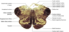
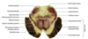
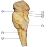
What are visible on the ventral surface of the medulla?
Prominent longitudinal columsn, the pyramid which run either side of the ventral median fissure.
Lateral to the pyramid lies an elongated elevation, the olive, within which lies the inferior olivary nucleus
Inferior olivary nucleus
This has connections primarily with the cerebellum and is involved in the control of movement.


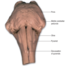
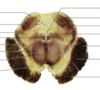
What delineates the transition from medulla to pons on the ventral surface of the brainstem?
The transverse system of fibres (the transverse pontine fibres) that originate from cells in the ventral pons and pass through the contralateral middle cerebellar peduncle to enter the cerebellar hemisphere. This massive system obscures the underlying pyramidal tracts
Features of the ventral surface of the midbrain?
Consists of a large column of descending fibres on either side, the crus cerebri or basis penduculi.
The two crura are separated by the interpeduncular fossa (basal cistern).
The crus cerebri is continuous with the internal capsule of the cerebral hemispherse and consists of corticobulbar and corticospinal fibres.
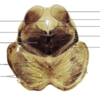
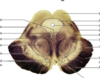
What portion of the brainstem is this?

Caudal part of the medulla

What happens to the pattern of grey and white matter as there is the transition from spinal cord to medulla?
The ventral horn becomes much more attenuated, the dorsal horn is replaced by the caudal part of the trigeminal sensory nucleus.
Trigeminal sensory nucleus
Regarded as the brainstem homologue of the dorsal horn as it receives afferent fibres conveying general sensation from the head, which enter the brainstem in the trigeminal nerve.
It is a large nucleus that extneds the whole length of the brainstem and into the upper segments of the spinal cord.
The caudal part is particularly associated with pain and temperature.
Where does the trigeminal nerve attach and what is the significance?
It attaches to the pons and therefore fibres that terminate in the parts of the trigeminal nucleus caudal to this level descend in the spinal tract of the trigeminal which lies immediately superficial to the nucleus
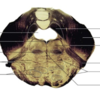
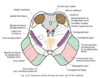
What portion of the medulla is this?

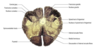
What are the features of the mid-medulla
Pyramids are prominent above their decussation ventrally.
Dorsally the ascending fibres of the dorsal columsn reach their termination in their respsective nuclei.
The dorsal columns consist of first-order sensory neurones with their cell bodies lying in the DRG of spinal nerves. They terminate in their nuclei upon the cell bodies of the second order neurones which course ventrally and medially, decussating as the internal arcuate fibres.
What happens to nerve fibres after the internal arcuate fibres
They turn rostraly, forming a distinct tract, the medial lemniscus, which runs through the rostral medulla, the pons and the midbrain to terminate on third-order neurones in the VPN of the thalamus