Vasculature of the CNS Flashcards
(55 cards)
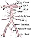
Arrangement of the arterial supply of the spinal cord
Three longitudinal vessels
Single anterior spinal artery
Paired posterior spinal artery
Composition of the anterior spinal artery
Arises in a Y shaped configuration from the two vertebral arteries at the level of the medulla and descends along the ventral suraface of the cord in the midline
Formation of the posterior spinal arteries
Arise either from the vertebral arteries or the PICA and run caudally on the posterolateral surface of the cord
Additional arterial supply of the spinal cord
Anterior and posterior spinal arteries alone are insufficient to supply the cord below cervical levels and receive serial reinforcement by anastomoses with radicular arteries derived from segmental vessels including the ascending cervical, intercostal and lumbar arteries
Passage of radicular arteries
Pass through the intervertebral foramina and divide into anterior and posterior branches which run with the dorsal and ventral spinal roots respsectively.
What is one particularly large radicular artetry?
Great radicular artery or artery of Adamkiewicz which may arise from a lateral intercostal or lumbar artery at any level between T8 and L3
Where is the blood supply of the spinal cord most vulnerable
In the thoracic region and in the anterior portion of the cord.
Occlusion of the anterior spinal artery leads to an acute thoracic cord syndrome with paraplegia and incontinence.
The spinothalamic modaliteis of pain and tempearture are preferentially lost with relative sparing of the dorsal columns
In what layer of spinal cord mater to the spinal arteries run?
Subarachnoid space
Venous drainage of the spinal cord
Follows basic pattern of arterial supply.
There are 6 longitudinal venous channels consisting primarily of anterior and posterior spinal veins which run in the midline.
There are sometimes incomplete/irregular bilaterally paired anterolateral and posterolateral veins situated near the sites of the ventral and dorsl nerve roots.
How do all of the veins of the spine drain
All drain via the anterior and posterior radicular veins into the internal vertebral venous plexus which is between the dura mater and vertebral periosteum.
With what does the internal venous plexus communicate
With an external venous plexus and thence with the ascending lumbar veins, the azygos and hemiazygos


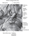
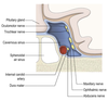
Entry of the ICA to the skull
Arises form the CCA and enters the middle fossa of the cranial cavity via the carotid canal.
Its course then follows a series of bends known as the carotid syphon, after which it passes through the cavernous sinus and then upwards on the medial aspect of the clinoid process, reaching the surface of the brain lateral to the optic chiasm.
What are the preterminal branches of the ICA?
Hypohpyseal
Opthalmic
Anterior choroidal
PComm
Hypophyseal arteries
Arise from the intra-cavernous section of the ICA to supply the neurohypophysis.
Also form the pituitary portal system.
Opthalmic artery
Passes into the orbit through the optic foramen.
Supplies the structures of the orbit, frontal and ethmoidal sinus, frontal part of scalp and dosrum of nose
Anterior choroidal artery
Supplies the optic tract, choroid plexus of lateral ventricle, hippocampus and some of the deep structures of the hemisphere including the internal capsule and globus pallidus
Calming voices make intra operative surgery pleasurable and almost memorable.
C: caroticotympanic artery (C2)
V: Vidian artery (C2)
M: meningohypophyseal trunk (C4)
I: inferolateral trunk (C4)
O: ophthalmic artery (C6)
S: superior hypophyseal artery (C6)
P: posterior communicating artery (C7)
A: anterior choroidal artery (C7)
A: anterior cerebal artery (C7)
M: middle cerebral artery (C7)
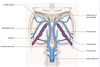
Terminal branches of the internal carotid
Lateral to the carotid arteries, the ICA branches into hte ACA and MCA.
Passage of the ACA
Courses medially above the optic nerve and passes into the great longiutdinal fissure between the frontal lobes of the cerebral hemispheres.
It is joined to the corresponding vessel of the other side by the AComm.
As it passes down the great longitudinal fissure, the ACA follows the dorsal curvature of the corpus callosum, branches ramifying over the medial surface of the frontal and parietal lobes.
Passage of the MCA
Passes laterally to enter the lateral fissure within which it subdivides with its branches supplying virtually the entire lateral surface of the frontal, parietal and temproal lobes.
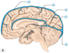
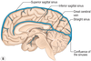
Territory supplied by the ACA
Includes motor and sensory cotrices for the lower limb.
Fine terminal branches also extend out of the great longiutdinal fissure to supply a anrrow lateral band of frontal and pariteral cortices.

Territories supplied by the MCA
PRimary motor and senosry cortices for the whole body excluding the lower limb.
Auditory and insula within the depths of the lateral fissure.