Cranial nerves and cranial nerve nuclei Flashcards

(101 cards)

1
Q
A
2
Q

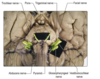
What are the afferent nuclei of the cranial nerves?

A

Trigeminal

Vestibular

Cochlear

Nucleus solitarius (visceral afferents)

3
Q

How can the efferent nuclei of the cranial nerves be divided?

A

On the basis of their embryological derivation, they can be divided into three groups, each lying in a discontinuous longitudinal column

4
Q

What are the three embryological columns of the efferent cranial nerves?

A

Somatic efferent cell column

Nuclei of the parasympathetic cell column

Nuclei of the branchimotor cell column

5
Q

Somatic efferent cell column

A

Lies near to the midline and consists of the nuclei of the III, IV, VI and XII nerves.

6
Q

Oculomotor nucleus

A

Lies in the ventral apex of the periaqueductal grey matter of the midbrain at the level of the superior colliculus.

Its efferent fibres run in the oculomotor nerve to innervate LPS and all of the extraocular muscles except SO and LR

7
Q

Trochlear nucleus

A

Midbrain at the ventral border of teh periaquedcutal grey matter, but at the level of the inferior colliculus.

Fibres leave in the trochlear nerve to innervate SO

8
Q

Abducens nuclei

A

Located in the caudal pons, beneath the floor of the fourth ventricle.

Innervates LR

9
Q

Hypoglossal nucleus

A

In the medulla

Innervates the intrinsic and extrinsic muscles of the tongue

10
Q

Which are the nuclei of the branchimotor cell column

A

Innervates striated muscles derived from the branchial arches

Trigeminal motor nucleus

Facial motor nucleus

Nucleus ambiguus

11
Q

Trigeminal motor nucleus

A

In the tegmentum of the mid-pons.

Supplies fibres to the trigeminal nerve which innervate the muscles of mastication, tensor tympani, tensor veli palatini, mylohyoid and anterior belly of digastric muscle

12
Q

Facial motor nucelus

A

In the caudal pontine tegmentum.

Innervates the muscles of facial expression and stapedius

13
Q

Nucleus ambiguus

A

Within the medulla

Sends motor fibres in the GPA, vagus and cranial part of the accessory nerve to innervate the muscles of the pharynx and larynx

14
Q

Nuclei of the parasympathetic column

A

Edinger-Westphal

Superior and inferior salivatory nuclei

Dorsal motor nucleus of the vagus

15
Q

Edinger Westphal

A

Most rostral cell group which lies in the midbrain periaqueductal grey matter adjacent to the oculomotor nucelus.

Its axons leave in the oculomotor nerve and pass into the cilairy ganglion from which postgnaglionic fibres innervate the sphincter pupillae and ciliary muscles within the eye

16
Q

Superior and inferior salivatory nulei

A

Lie in the pontine tegmentum

17
Q

Superior salivatory nucleus

A

Supplies preganglionic fibres to the facial nerve that terminate in the pterygopalatine and submandibular ganglia.

Postganglionic fibres innervate the lacrimal gland nasal and oral mucous membranes.

Those from the submandibular innervate the submandibular and sublingual salivary glands

18
Q

Inerior salivatory nuclei

A

Sends preganglionic fibres into GPA which termiante in the otic ganglion, which in turn sends postganglionic axons to the parotid gland

19
Q

Dorsal motor nucleus of the vagus

A

Medulla

Rostral portion lies immediately beneath the floor of the fourth ventricle, lateral to hypoglossal.

Fibres leave in the vagus nerve and are widely distributed to thoracic and abdominal viscera

20
Q

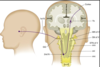
Contents of the oculomotor nerve

A

Somatoic motor axons that innervate the extraocular muscles.

Also contains preganglionic parasympathetic neruoens that control the smooth muscle of the eye via the ciliary ganglion

21
Q

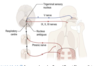
Passage of oculomotor neurones in the brainstem.

A

Oculomotor nucleus and Edinger Westphal.

Course ventrally through the midbrain tegmentum, with many of them traversing the red nucleus to exit on the medial aspect of the crus cerebri within the interpeduncular fossa

22
Q

At what site does the oculomotor nerve leave the midbrain?

A

On the medial aspect of the crus cerebri within the interpeduncular fossa, passing between PCA and SCA

23
Q

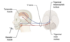
Oculomotor course after leaving the brainstem

A

Runs anteriorly, lying in the wall of the cavernous sinus, before gaining access to the orbit through the superior orbital fissure.

24
Q

Somatic motor function of oculomotor

A

Elevates, depresses and adducts the eyeball.

Elevates upper eyelid.

25
Course of parasympathetic fibres of III
EW-\> ciliary ganglion located within the orbit behind the eyeball. Postganglionic neurones run in short ciliary nerves to innervate the sphincter pupillae muscle of the iris and the ciliary muscle contained within the cilliary body.
26
Light reflex.
Small contingent of optic tract fibres that pass directly from the eye to the pretectal area, rostral to the superior colliculus, rather than to the LGN. Neurones project bilaterally to the EW nuclei, from which efferent fibres leave in the oculomotor nerve.
27
Accommodation reflex
Fixation upon a nearby object, by convergence of the optic axes, involves concomitant contraction of the ciliary muscles to increase the convexity of the lens and focus the image. It is also accompanied by pupillary constriction. Involves the visual cortex, with corticobulbar fibres activating the PNS of the EW nuclei bilaterally
28
Passage of trochlear nerve fibres in the brainstem
Arise inthe trochlear nucleus in the ventral part of the midbrain periaqueductal grey matter, at the level of the inferior colliculus. The efferent axons pass dorsally, around the periaqueductal grey and decussate in the midline. It emerges from the dorsal aspect of the brainstem, caudal to the inferior colliculus
29
Passage of trochlear after leaving the brainstem
Leaves caudal to the inferior colliculus. Coursing around the cerebral peduncle to gain the ventral aspect of the brain, passing between the posterior cerebral and superior cerebellar arteries. Runs anteriorly lying in the lateral wall of the cavernous sinus entering the orbit through the SOF.
30
Action of SO
Most importantly, with the eye adducted, it depresses the visual axis.
31
32
33
Passage of abducens within the brain stem
Abduens nucleus which lies below the floor of the fourth ventricle in the caudal pons. Efferent axons course ventrally through the pons and emerge at the junction between the pons and the pyramid of the medulla
34
Passage of abducens after leaving the pons
Passes anteriorly through the cavernous sinus to enter the orbit through the SOF
35
36
Internuclear opthalmoplegia
Classically in MS due to demylination of the MLF in the brainstem which interferes with conjugate ocular deviation. Typically on horizontal gaze, the abducting eye moves normally but the adducting eye fails to follow. Adduciton is preserved on convergence.
37
Roots of the trigeminal
Two adjacent roots, a large sensory and a smaller motor root on the ventrolateral aspect of the pons where it merges with the middle cerebellar peduncle.
38
Sensory component of the trigeminal
Primary sensory neurones with peripheral processes distributied via V1-3. Sensations of touch, pressure, pain and tempearture are relayed from the face and scalp, cornea and oral cavities including the teeth and gums and the paranasal sinsues. It also innervates the intracranial dura mater and the intracranial arteries. Proprioceptive fibres are carried from the muscles of mastication and the TMJ
39
Location of the cell bodies of afferents of the trigeminal?
Located in the trigeminal (or semilunar) ganglion, located at the convergene of the ophthalmic, maxillary and mandibular nerves. This excludes the proprioceptive fibres. The central processes of these cells terminate in the trigeminal sensory nucleus.
40
Arrangement of the trigeminal sensory nucleus
Large nucleus that extends throughout the length of the brainstem and into the upper cervical SC. Consists of three subnuclei- chief sensory nucleus, mesencephalic nucleus, spinal nucleus
41
Chief sensory nucleus
Lies in the pontine tegmentum, close to the entry of the trigeminal nerve
42
Mesencephalic nucleus
Extends rostrally into the midbrain
43
Spinal nucleus of trigeminal
Extends caudally through the medulla into the cord, where it becomes continuous wit the substantia gelatinosa.
44
Segregated distribution of afferent fibre termination in the trigeminal nucleus,
Depends on the modality being served. Touch and pressure fibres terminate in the principal nucleus. Pain and temperature end in the spinal tract of the trigeminal. Proprioceptive afferents from the muscles of mastication and the TMJ have their cell bodies in the mesencephalic nucleus (rather than the trigeminal ganglion).
45
What are the only primary afferents to have their cell bodies located in the CNS?
Propriorceptive afferents of the trigeminal nerve, with their nuclei in the mesencephalic nucleus.
46
Second order neurones of the trigeminal.
Decussate to form the contralateral trigeminothalamic tract (trigeminal lemniscus) which terminates in the contralateral VPN of the thalamus, which in turn sends fibres to the postcentral gyrus of the parietal lobe.
47
Reflexes mediated by trigeminal?
Corneal Sneeze and cough Jaw jerk
48
Corneal reflex
Afferent limb V Efferent VII
49
Sneeze and cough reflexes
Mediated by trigeminal afferents ending in the trigeminal sensory nucleus. Indirect connections are made with the diaphragmatic, intercostal and abdominal muscle motorneurones and with the nucleus ambiguus
50
Jaw jerk reflex
Afferent and efferent are V, to the trigeminal mesencephalic nucleus
51
Course of the motor component of trigeminal
Arise from cells in the trigeminal motor nucleus inthe pontine tegmentum, medial to the principal sensory nucleus. Axons leave the pons in the motor root of the trigeminal and then join the mandibular divsion of the nerve.
52
Muscles innervated by trigeminal
Mastication (masseter, temproalis, lateral and medial pterygoids) Tensor tympani Tensor veli palatini Mylohyoid Anterior belly of digastric
53
Location of the facial nerve
Ventrolateral aspet of the caudal pons, near the pontomedullary junction in a rergion known as the cerebellopontine angle
54
Roots of the facial nerve
Lateral nervus intermedius- sensory and parasympathetic Medial- motor
55
Sensory fibres of the facial nerve
Taste sensation from the anterior two thirds of the tongue, floor of the mouth and palate and also cutaneous senation from part of the external ear.
56
Where are the cell bodies of primary afferents of the facial nerve?
Geniculate ganglion within the facial canal of the petrous temporal bone.
57
Where do the central processes of facial taste fibres terminate?
Rostral part of the nucleus solitarius of the medulla.
58
Passage of ascending fibres from the nucleus solitarius
Project to VPN of the thalamus, which in turn sends sensory fibres to the postcentral gyrus of the parietal lobe. Afferent fibres that carry cutaneous sensation terminate in the trigeminal nucleus.
59
`passage of motor fibres of the facial nerve
Facial motor nucelus of the caudal pontine tegmentum. Axons initially pass dorsally, looping over the abducens nucleus beneath the floor of the fourth ventricle, leaving the brainstem in the motor root of the facial nerve.
60
61
Reflexive proection of the eye and the facial nerve
Corneal reflex through fibres from the superior colliclus and the trigeminal sensory nucleus.
62
Reflex contraction of stapedius
Fibres from the superior olivary nucleus, part of the central auditory pathway stimulate reflex stapedius contraction
63
Corticobulbar innervation of facial muscles.
Corticobulbar fibres that control motor neurones supplying the upper face muscles are distributed bilaterally. Those that control the motor neurones supplying the muscles of the lower face are entirely crossed. Unilateral upper motor neurone lesiosn therefore give rise to paralysis fo the lower facial muscles.
64
Passage of parasympathetic fibres of the facial nerve?
Superior salivatory nucleus of the pons. Leave the brainstem in the sensory root of the facial nerve (nervus intermedius), pass to the parasympathetic ganglia: the submandibular and pterygopalatine where they synapse with postganglionic neurones
65
Submandibular ganglion
Innervates the submandibular and sublingual salivary glands
66
Pterygopalatine ganglion
Innervates the lacrimal gland and the nasal and oral mucous membranes
67
What are the two component parts of the vestibulocochlear nerve?
Vestibular nerve Cochlear nerve
68
Where does the vestibulocochlear nerve attach to the brainstem?
At the junction of the medulla and pons at the CPA
69
Path of the vestibular nerve
Fibres make dendritic contact with hair cells of the membranous labyrinth with their cell bodies located within the vestibular ganglion within the internal auditory meatus. The central processes mostly end in the vestibular nuclei of the rostral medulla (there are four such nuclei- superior, inferior, medial and lateral) which are located close together between the lateral part of the floor of the fourth ventricle
70
Deiter's nucleus
Lateral vestibular nucleus
71
What happens to fibres from the lateral vestibular nucleus?
Descend ipsilaterally in the lateral vestibulospinal tract
72
What other structure takes contributions from the vestibular fibres?
The MLF
73
MLF
Extends throughout the brainstem and into the SC Its descending component is also known as the medial vestibulospinal tract The ascending component establishes connection with the nuclei of the abducens, trochlear and oculomotor nerves to coordinate the movement of the head and eye movements.
74
To what other portions of the brain do vestibular nuclei efferents project?
Project to the flocculonodular lobe of the cerebellum which is concerned with the control of equilibirum Also ascend to the contralateral VPN which in turn projects to the cerebral cortex.
75
Passage of the cochlear nerve
Fibres make dendiritic contact with hair cells of the organ of Corti within the cochlear duct of the inner ear. The cell bodies lie within the cochlea and are collectively called the spiral ganglion. It joins the brainstem at the level of the rostral medulla. Its fibres bifurcate and end in the dorsal and ventral cochlear nuclei, close to the inferior cerebellar particle. There is a complicated ascending auditory pathway to the thalamus and cerebral cortex
76
Describe the ascending auditory pathway to the level of the inferior colliculus.
From the cochlear nuclei, second order neurones ascend into the pons, some of them crossing to the other side of the pontine tegementum as the trapezoid body. Some of these fibres may terminate in the superior olivary nucleus which is the site of olivocochlear fibres which leave the brainstem in VIII and end in the organ of corti. From the superior olivary nuclei, ascending fibres comprise the lateral lemniscus which runs through the pontine tegmentum to end in the inferior colliculus. Some of these neurones terminate in a small pontine nucleus, the nucleus of the lateral lemniscus.
77
Olivocochlear fibres
Run from the superior olivary nucleus in CN 8 to the organ of Corti, are though to have an inhibitory function and modulate the transmission of auditory information to the cochlear nerve.
78
Action of the superior olivary nucleus and nucleus of the lateral lemniscus
Establish reflex connections with motor neurones of the trigeminal and facial motor nuclei mediating contraction of the tensor tympani and stapedius.
79
Ascending auditory pathway from the inferior colliculus
Sends axons to the MGN of the thalamus. Axons from the MGN pass through the internal capsule to the primary auditory cortex in Heschel's gyrus (transverse temporal gyrus of Heschl) There is a tonotopical representation of the cochlea which is analogous to somatotopic sensation for patthways of general sensation. Within the brainstem, some fibres decussate and others do not, central ascending auditory pathway lesions therefore do not classically cause monaural deafness but are rather manifest as a loss of auditory acuity and inability to localise the directional origin of sounds
80
81
Region of temproal lobe surrounding the primary auditory cortex?
Auditory association cortex (Wernicke's area)
82
Attachment of GPA to the brainstem
Linear series of small rootlets, lateral to the olive in the rostral medulla
83
84
From where do afferent fibres of the glossopharyngeal nerve convey information
Receptors for sensation in the pharynx, posterior third of the tongue, Eustachian tube and middle ear Taste buds of the pharynx and posterior third of the tognue Chemoreceptors in the carotid body and baroreceptors int he carotid sinus
85
Where do glossopharyngeal somatic afferents end in the brainstem
In the trigeminal sensory nucleus
86
Where do visceral and taste fibres of the glossopharyngeal nerve terminate
In the nucleus solitarius of the medulla.
87
What is the motor function of the glossophrayngeal
Innervates stylopharyngeus Sends preganglionic parasymthpathetic fibres from the inferior salivary nucleus of the rostral medulla to the otic ganglion, which in turn innervates the parotid sensory gland
88
Afferent input of the vagus
Receptors for general sensation in the pharynx, larynx, oesophagus, tympanic membrane, external auditrory meatus and part of the concha of the middle ear. Chemoreceptors in the aortic bodies and baroreceptors int he aortic arch, Receptors from the thoracic and abdominal viscera
89
Where do afferents carying general sensation from X end?
In the trigeminal sensory nucleus
90
Where do afferents carying visceral information from X end?
In the nucleus solitarius
91
Where do the somatic motor fibres of the vagus arise?
From the nucleus ambiguus of the medulla. They innervate the muscles of the soft palate, pharynx, larynx and upper part of the oesophagus.
92
By convention, where do most caudal efferents from the nucleus ambiguus leave the brainstem?
Leave the brainstem in the cranial roots of the accessory nerve but transfer to the vagus proper at the jugular formaen.
93
Origin of parasympthatic vagal fibres
From the dorsal motor nucleus of the vagus, in the medulla immediately beneath the fourth ventricle
94
Outline the cranial root of the accessory nerve
Emerges from the lateral aspect of the medulla as a linear series of rootlets, immediately caudal to the vagus nerve. It carries fibres that have their origin in the caudal part of the nucleus ambiguus of the medulla. At the level of the foramen, these join the vagus nerve and are distributed with it to the muscles of the soft palate, pharynx and larynx
95
Spinal roots of the accessory nerve
Arise from motor nuerones in the ventral horn of spinal grey matter at C1-5. leave the cord not though the ventral roots but via a series of rootlets that emerge from the lateral aspect of the cord, midway between the dorsal and vetnral roots. These course rostrally, entering the cranium through the foramen magnum. At the medulla, the spinal root of the accessory nerve is joined briefly by the cranial rootlets but separate as the nerve leaves the cranial cavity through the jugular foramen. Here the fibres of the cranial root join the vagus. The fibres of the spinal root pass to the sternomastoid and trapezius
96
Action of the hypogloosal
Innervates both the intrinsic and extrinsic muscles of the tongue
97
Passage of hypoglossal nerve fibres
Originates in the hypoglossal nucleus, immediately beneath the floor of the fourth ventricle near the midline. Axons course ventrally through the mdulla and emerge from its ventrolateral aspect as a linear seires of rootlets between the pyramid and olive. It receives afferents from the nucleus solitarius and trigeminal sesnory nucleus. These are involved in the reflex movements. It also receives corticobulbar fibres.
98
What is the only tongue muscle not innervated by XII?
Palatoglossus (X)
99
Dysphonia and depression of gag reflex Unilateral wasting of sternomastoid and trapezius. Unilateral weakness, wasting and fasciculation of the tongue?
Jugular foramen syndrome
100
Question 1 The eye moves to stabilize the gaze or to track movement of objects. Eye movements are controlled by the extraocular muscles. What is the function of the superior rectus eye muscle? Adducts the eye Depresses the eye after abduction Depresses the eye after adduction Elevates the eye after abduction Elevates the eye after adduction
Innervation and actions of extraocular muscles: Complimentary muscle in other eye= inferior oblique
101
Perception of the direction and characteristics of sound have to be conducted to the primary auditory cortex in the brain. The auditory nerve impulses in the acoustic nerve pass through which of the following to reach the primary auditory cortex? Contralateral cochlear nuclei in the medulla Ipsilateral vestibular nuclei Ipsilateral lateral geniculate body Contralateral medial geniculate body Ipsilateral superior colliculus
Contralateral MGN