Cerebellum Flashcards
(31 cards)


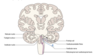
Superior surface of the cerebellum
Lies beneath the dural tentorium cerebelli with the superior vermis raised, forming a midline ridge.
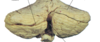
Inferior vermis
Lies in a deep groove between the hemispheres.
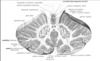
Folia
Folds of the surface of the cerebellum which are orientated approximately trasnveresely.
Between the folia lie fissures of varying depths, some of which can be used to divide the cerebellum anatomically into theree lobes
Primary fissure
Separates anterior lobe from the much larger posterior lobe.
Posterolateral fissure
Demarcates the location of the flocculus and the bermis, which together form the flocullonodular lobe


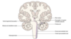
Arrangement of the white matter of the cerebellum
Made up largely of afferent and efferent fibres that run to and from the cortex, towards which it extends characteristic irregular branch like projections, arborvitae.
Buried deep within the white matter are four bilaterally paired cerebellar nuclei.
What are the four white matter nuclei of the cerebellum?
Dentate
Fastigial
Emobliform
Globose


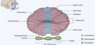
What is the histological arrangement of the cerebellar cortex?
3 layers
Molecular- fibre rich
Intermediate- Purkinje cell layer
Granular layer
What are the principle afferent projections to the cerebellum
Spinocerebellar (from SC)
Olivocerebellar (inferior olivary nucleus)
Vestibulocerebellar (vestibular nuclei)
Pontocerebellar (pons)
Passage of afferent fibres to the cerebellum
Mostly termiante in the cerbellar cortex, where they are excitatory tocortical neurones.
Fibres enter the cerebellum through one of the peduncles as eiether mossy fibres of climbing fibres depending on their origin.
Afferents from the inferior olivary nuclei end up as ?
Climbing fibres
All other afferent fibres end up as
Mossy fibres.
What hapens to mossy fibres in the cerebellar cortex
Branch to supply several folia and end in the granular layer, in synaptic oncat with granule cells.
The axons of granule cells pass towards the surface ofr the cortex and enter the molecular layer.
Here they bifurcate to produce two parallel fibres that are originated along the axis of the folium

Outline the Purkinje layer of the cerebellar cortex
A unicellular layer of the somata of Purkinje neurones.
Their profuse dendritic arborisations extend into the cortex, into the molecular layer.
The arborisations are flattened and orientated at right angles to the long axis of the folium. They are therefore traversed by numerous parallel fibres.
Inhibitory modulation of intracortical circuitry is provided by other neurones such as Golgi, basket and stellate cells.
most of these fibres do not leave the cerebellum and terminate in the deep cerebellar nuclei.
Climbing fibres
Originate from the inferior olivary nucleus of the medulla.
Provide relatively discrete excitatory input to Purkinje cells.
Axon collaterals of climbing fibres ecite the nuroens of the deep cerebellar nuclei.
Purkinje cells are GABAergic, which means tat the output of the whole cerebellar cortex is mediated through inhibition of cells in the cerebellar nuclei
Cerebellar nuclei from medial to lateral
FGED
Fastigial
Globose
Emboliform
Dentate
Dentate nucleus
Thin layer of nerve cells reminiscent of a crinkled bag.