Cerebral Hemisphere and Cerebral Cortex Flashcards
(114 cards)
1
Q
What proprtion of the surface area of the cortex is hidden within the depths of sulci
A
70%
2
Q
Corona radiata
A
Radiation of fibres between the cortical surface and the internal capsule
3
Q
A
4
Q

A

5
Q
A
6
Q
A

7
Q

A

8
Q

A
9
Q

A

10
Q
A

11
Q
A
12
Q

A

13
Q

A

14
Q

A
15
Q
A

16
Q

A

17
Q
A
18
Q

A
19
Q

A

20
Q

A
21
Q

A
22
Q

A
23
Q

A
24
Q
A
25

26

27

28
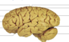
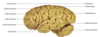
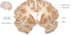
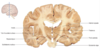
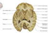
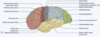
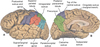
What are the lobes of the brain?
Frontal
Parietal
Temporal
Occipital
With the two hemispheres separated by the longitudinal fissure
29
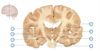
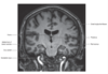
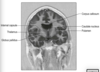
What is found at the base of the larteral (Sylvian) fissure
Insula
30
What are the parts of the frontal, temporal and parietal lobes that overlies the insula?
The opercula
31

32


33
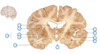
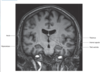
What constitutes the frontal lobe
The entire region in front of the central sulcus.
Immediately in front is the precentral gyrus (primary motor cortex)
In front of the precentral gyrus, the frontal lobe consists of a more variable pattern of convolutions, of which the superior, middle and inferior frontal gyri
34
What is found posterior to the central sulcus
The parietal lobe, the most anterior part of which is the postcentral gyrus, the primary somatosensory cortex.
Behind the postcentral gyrus, on the lateral hemisphere, the intraparietal sulcus divides the rest of the parietal lobe into superior and inferior parietal lobules
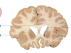
35
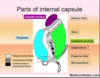
What is the boundary between the parietal and occipital lobes
It is clearly marked on the medial surface by the deep parieto-occipital sulcus
36
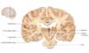
What are the important landmarks of the occipital cortex
Does not bear significant landmarks on lateral surface, on the medial surface the prominent calcarine sulcus indicates the locaiton of the primary visual cortex
37
Arrangement of the temporal lobe
On the lateral surface, the temporal lobe is divided into three principle gyri that run roughly parallel to the lateral fissure, the superior, middle and inferior temporal gyrus,
The superior temporal gyrus includes the primary auditory cortex, most of this functional region is situated on the superior bank of the gyrus within the lateral fissure on the transverse temporal gyri of Heschel

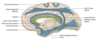
38
What curves around the corpus callosum?
The cingulate gyrus, separated from the rest of the hemisphere by the cingulate sulcus.
39
With what is the cingulate gyrus continuous?
With the parahippocampal gyrus of the temporal lobe
40
What are the regions of the cerebral cortex with archicortex and paleocortex?
Hippocampus
Other parts of the temporal lobe
Olfactory system
41
Layers of neocortex
Molecular
External granular
External pyramidal
Interanl granular
Internal pyramidal
Multiform
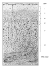
42
Molecular cell layer
Few cell bodies, many dendritic and axonal processes in synaptic interaction
43
External granular layer
Contains many small neurones which establish intracortical connections, receiving local afferent inputs
44
External pyramidal layer
Medium sized neurones giving rise to commissural fibres
45
Internal granular layer
Site of termination of afferent fibres from the specific thalamic nuclei
46
Internal pyramidal layer
Origin of projection fibres to extracortical targets such as the BG, thalamus, brainstem and SC.
In the primary motor cortex of the frontal lobe this layer contanis the giant Betz cells which project fibres into the pyramidal tract
47
Multiform layer
Contains mixture of association and projection neurones
48
Posterior part of the cerebrum
Recievs sensory input from the outisde world in the somatosensory, occpital and temporal regions
49
Cortical zones adjacent to sensory regios
Information is elaboraed to permit identificatoin of objects by touch, sight and hearing in a modality specific perception.
These areas of association cortex are critical for multimodal and spatial recognition of the environment
50
Limbic systems
Enable storage and retrieval of information processed in the posterior hemispheric regions
51
Anterior part of the cerebrum
Concerned with the organisation of mvoement and the strategic guidance of complex motor behaviour over time
52
Primary motor cortex= Brodman area
4
53
What proportion of corticospinal and corticobulbar fibres arise from the primary motor cortex?
30%
54
Principle afferents into the primary motor cortex
VLN which in turn receives input from the dentate nucleus of the cerebellum and from the globus pallidus of the BG
55
Premotor cortex= Brodmann ara
6
56
Featurs of the premotor cortex
On the lateral surface of the hemisphere, this includes the posterior portions of the superior, middle and inferior frtonal gyri.
On the medial surface of the hemisphere the premotor cortex includes a region referred to as the supplementary motor cortex.
57
Features of supplementary motor cortex
Has somatotopic representation of the body which appears to be bilateral in both hemispheres, in contrast to the priamry motor cortex
58
What is the effet of stimulation of premotor cortical areas
Induces movements that are less focused than those elicited fromt he priamry motor cortex and that involve groups of functionally related muslces.
e.g. postural in nature.
Premotor cortical areas are thought to funciton in the programming of and preparation for movement and in the control of posture.
59
What is the principal subcoritcal input to premotor cortical regions?
Ventral anterior nucleus of the thalamus.
This in turn receives fibres from the globus pallidus and SN
60
What is found in the middle frontal gyrus?
Frontal eye field
61
Brodman area 8
Frontal eye field
62
Function of frontal eye field
Region controls voluntary conjugate deviation of the eyes.
Unilateral damage to this area causes conjugate deviation of the eyes towards the side of the lesion
63
What is found in the inferior frontal gyrus?
Broca's area- motor speech
64
Brodman's area 44 and 45

Broca's area
65
What is found anterior to premotor areas?
Preforntal cortex
66
Connections of the prefrontal cortex
Rich connections with parietal, temporal and occipital cortex through long association fibres running in the subcortical white matter.
67
Function of the prefrontal cortex?
Cognitive functions of high order including intellectual, judgemental and predictive faculties and the planning of behaviour
68
What is the most anterior part of the parietal lobe?
Postcentral gyrus, running parallel to the central sulcus.
Functionally this area is the primary somatosensory cortex
69
BRodmann's areas 1 and 2
Priamry somatosensory cortex
70
Where do thalamocortical nuerones terminate?
In the primary somatosensory cortex. Thalmic origin of VPN (which in tur receives input from the medial lemnisus, spinal lemnisucs, spinothalamic tract and trigeminothalamic tracts.
71
What is found posterior to the postcentral gyrus?
The association cortex.
72
Superior parietal lobule is responsible for?
Interpretation of general sensory information and for conscoius awareness of the contralateral half of the body.
Lesions here impair the interpretation and understanding of sensory input and may cause negelct of the opposite side of the body.
73
Inferior parietal lobule
Interfaces between the somatosensory cortex and the visual and auditory association cortices of the occipital and temporal lobes.
in the dominant hemisphere it contributes to language functions
74
Divisions of the lateral surface of the temporal lobe?
Superior
Middle
Inferior temporal gyri.
75
Location of the primary auditory cortex
In the superior temporal gyrus.
Lies in the superior bank of the gyrus, normally hidden within the lateral fissure.
Its precise location is marked by the small transverse temporal gyri or Heschl's convolutions
76
Location of the auditory association cortex
Lies surrounding and immediately posterior to the primary auditory cortex.
In the dominant hemisphere, this region is known as Wernicke's area.
It is crucial for understanding the spoken word.
77
Location of the hippocampus
Inferomedial part of the temporal lobe.
Lies in the floor of the inferior horn of the lateral ventricle, deep to the parahippocampal gyrus.
78
Location of the amygdala
Anterior end of the hippocampus and temproal pole.
Receives fibres from the olfatctory tract
79
What marks the boundary between the occipital and parietal lobes?
The deep parieto-occipital sulcus
80
What is found on the medial surface of the occipital lobe?
The priamry visual cortex on the calcarine sulcus. It is immediately above and below the sulcus, much of it hidden int he depths of the sulci.
81
Cortical representation of the visual field
Each lateral half of the visual field is represented in the priamry visual cortex of the contralateral hemisphere.
The upper half of the visual field is represented below the calcarine sulcues and the lower half above.
82
Visual association cortex
Constitutes the rest of the occipital lobe.
It is concerned with the interpretation of visual images.
Damage to this region causes disorders of visual interpretation and recognition

83
What provides interface between the auditory and visual association areas/
The angular gyrus and supramarginal gyrus
84
What are the three categroies of white matter fibres?
Association
Commissural
Projection
85
Association fibres
Interconnect cortical sites within one cerebral hemisphere
86
Commissural fibres
Run from one hemisphere to the other
87
Projection fibres
Pass between the cerebral cortex and subcortical structures.
88
SLF
Interconnects the frontal and occiptial lobes.
A subsidiary of this bundle, the arcuate fasciculus, links gyri int he frontal and temporal lobes, which are important for language function
89
ILF
Runs from the occipital to temporal poles and contributes to the function of visual recognition
90
Uncinate fasciculus
Connects the anterior and inferior parts of the frotnal lobe with the temproal gyrus.
91
Cingulumn
Lies within the cingulate gyrus and courses around the corpus callosum, connecting the frontal and parietal lobes with the parahippocampal and adjacent temporal gyri.
92
Prosopagnosia
INabiltiy to identify individual faces (can occur through ILF damage)
93
What are the major interhemispheric commisural fibres?
Anterior commissure
Posterior commissure
Corpus callosum
Hippocampal commissure
94


95

96

97


98


99


100
Parts of the corpus callosum
Rostrum
Genu
Body
Isthmus
Splenium
101
Forceps minor
Anteriorly
102
Forceps major
Posteriorly
103
Location of the anterior commissure
Runs transversely in front of the anterior column of the fornix and interconnects the inferior and middle temproal gyri and the olfactory regions of the two sides
104
Hippocampal commissure
Consists of transverse fibres linking the posterior columns of the fornix on each side
105
Corona radiata
Expansive radial distribution of projection fibres within the cortex
106
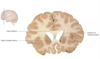
Location of the internal capsule
Between the thalamus and caudate nucleus medially and the lentiform nucleus laterally.
107
Arrangement of the internal capsule
Anterior limb
Genu
Posterior Limb
Retrolenticular part
108
Connections of the anterior limb of the internal capsule
Connections between the mediodorsal nucleus of the thalamus and the prefrontal cortex and also frontopontine fibres that project to the pontine nuclei in the basal portion of the pons
109
Connections of the posterior limb of the internal capsule
Contains corticobulbar and corticospinal motor fibres.
Also contains thalamocortical projections passing from the VPN to the primary somatosensory cortex and from the vetnral anterior and ventral lateral nuclei to motor regions of the frontal lobe
110
Components of the retrolenticular part of the internal capsule
Consists of fibres arising from the MGN and LGN of the thalamus that pass to the auditory and visual cortices as the auditory and visual radiations.

111
Meyer's loop
Thalamocortical fibres that loop forwards over the inferior horn of the lateral ventricle.
112
A large part of the brain is concerned with language.
Which of the following are features of Wernicke’s area?
It is usually found in the right superior temporal gyrus
Its main function is to express meaning
Lesions in this area result in dysarthria
It is connected to Broca’s area by the arcuate fasciculus
It is usually damaged by vascular occlusion of the left anterior cerebral artery
About 90% of the population are left hemisphere/right hand dominant. Amongst these people, Wernicke’s area is found in the left superior temporal gyrus. Its main function is to understand speech. Hence lesions in the area result in receptive aphasia, and patients have difficulty in understanding the speech of others, retrieving the appropriate names of objects, and monitoring their own speech, although they might appear to speak fluently and they may not be aware of the gross errors in their speech. Broca’s area is usually found in the left inferior frontal gyrus. Its main function is to express what the subject wishes to say. Hence a patient with a lesion in Broca’s area understands other people’s speech. However, their own speech is slow, hesitant and telegraphic. Patients are well aware of their speech problem. The two speech areas are supplied by branches of the left middle cerebral artery. Broca’s and Wernicke’s areas are connected by the arcuate fasciculus.
113
What is often associated with a stroke in the internal capsule of the left cerebral hemisphere?
Paralyis or paresis of the left limbs
A left-sided homonymous hemianopia
Dysarthria
Blockage of the basilar artery
Tremor at rest
The left cerebral hemisphere is usually dominant for speech function. A lesion in the internal capsule on that side, therefore, may interrupt descending cortical efferent fibres from the cerebral cortex to the cranial nerve nuclei involved in articulation.
114
