Spinal Cord Flashcards

(101 cards)

1
Q
A
2
Q

What are the two enlargements of the spinal cord?

A

Cervical and lumbar

3
Q

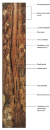
Cervical enlargement

A

Consists of cord segments C4-T1 and provides innervation for the upper limb via the brachial plexus.

4
Q

Lumbar enlargement

A

Made up of L1 to S3 and is associated with innervation of the lower limb via the lumbosacral plexus

5
Q

Conical termination of the spinal cord?

A

Conus medullaris

6
Q

What extends from the tip of the conus medullaris?

A

The filum terminale which extends caudally and is attached to the dorsal suface of the first coccygeal vertebra

7
Q
A
8
Q

From what point onwards does the spinal column no longer occupy the entire length of the SC?

A

Third month.

Thereafter the rate of elongation of the vertebral column exceeds that of the spinal cord.

As a result in adult life it terminates at L1/2

9
Q

Relationship of SC segments to their spinous processes cervical region

A

C spine segments lie around 1 spine higher than their corresponding vertebra

10
Q

Relationship of SC segments to their spinous processes thoracic region

A

2 spines higher

11
Q

Relationship of SC segmnets to their spinous processes lumbar region

A

4 spines higher

12
Q

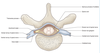
Initial formation of spinal nerves

A

Originate as two linear series of nerve fasicles attached to the dorsolateral and ventrolateral aspects of the cord.

These coalesce to form dorsal or ventral roots

13
Q

Passage of the spinal roots

A

Pass to their corresponding intervetrebral foramen in or near which they join to form the spinal nerve proper

14
Q

Contents of the dorsal spinal roots

A

Primary afferent neurones.

Nerve cell bodies located in the DRG.

15
Q

Contents of the ventral root

A

Efferent motor fibres

16
Q

What happens to the spinal nerves immediately after leaving the intervertebral foramina?

A

Divide into dorsal (posterior) and ventral (anterior) rami

17
Q

Dorsal spinal rami

A

Supply the muscles and skin of the back

18
Q

Ventral spinal rami

A

Supplies the muscles and skin on the front of the body and also the limbs

19
Q

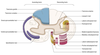
Denticulate ligament

A

Found midway between the dorsal and ventral roots of the spinal nerves.

It is a flat, membranous continuation of the pia.

It has a free lateral border for much of its length but it intermittently has lateral projections that tether the SC to the arachnoid and through it to the dura

20
Q

What space separates the spinal cord dura from the bony wall of the vertebral canal?

A

Epidural space

21
Q

To what level do the spinal arachnoid and dura continue?

A

S2

22
Q

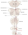
Division of the spinal cord

A

Divided into two symmetrical halves by a dorsal median sulcus and ventral median fissure

23
Q

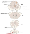
Why are there changes in the relative configuration of white and grey matter in the SC at different levels

A

Higher levels contain greater amounts of white matter as the ascending tracts gain fibres at each successive level

24
Q
A
25
Arrangement of the grey matter of the spinal cord
Can be divided on the basis of cytoarchitecture into ten zones, known as Rexed's laminae, which are numbered sequentially from dorsal to ventral
26
How may afferent fibres travel in the spinal cord.
They mostly terminate near their point of entry but may travel for varying distances in either direction, running in the dorsolateral fasciulus or Lissauer's tract which is located superficial to the tip of the dorsal horn. They may therefore establish connections over several segments of spinal grey matter.
27
Where do cutaneous dorsal root fibres tend to terminate
In the superifical laminae
28
Where do proprioecptive nerve root fibres terminate?
In the deeper laminae
29
Substantia gelatinosa
Tip of the dorsal horn approximating to Rexed's laminae I-III. Receives collaterals of the smallest diameter myelinated (group A delta) and unmyelinated (group C) afferents that are associated with nociception
30
What occurs in the substantia gelatinosa
Complex interactions with other types of afferent terminal interneurones and with descending pathways from the brain which control the transmission of pain information to ascending spinothalamic and spinorteicular tract neurones throughout the dorsal horn. i.e. Gate theory.
31
Basic mechanics of gate theory.
Large diameter afferents excite the dorsal horn interneurones which use enodengous encephalin (opioid peptide) as a neurotransmitter. These interneurones establish presynaptic contacts with the terminals of nociceptive primary afferent neurones. They presynaptically inhibit these neurones, thus decreasing the release of transmitter from afferent terminals and the subsequent excitation of ascending tract neurones.
32
Descending modulation of afferent signalling.
Descending serotonergic fibres from the nucleus raphe magnus of the medulla nd noradrenergic fibres of the locus coeruleus of the pons can excite these enkephalinergic dorsal horn interneurones, inhibiting the onward tranmission of nociceptive encephalin.
33
Clarke's column
Found in Rexeds VII in cord levels C8-L3. Cell bodies which are the origin of ascending fibres of the dorsal spinocerebellar tract. The cells of Clarke's column receive afferent input from mechanorecepetors
34
Contents of Rexed's lamina VII
C8-L3, Clarke's bodies At the thoracic and upper lumbar levles, its lateral parts also contain preganglionic sympathetic neurones that constitute the lateral horn, while at the sacral levels it contains preganglionic parasympathetic neurones
35
Composition of lamina IX
Corresponds to groups of motor neurones that innervate skeletal muscle. Alpha motor neurones (extrafusal) Gamma motor neurones (intrafusal)
36
General arragnement of motor neurones in the cord
Neurones innervating axial musculature tend to be located medially, whilst those innervating limb muscles are found more laterally.
37
What is found in the ventral cord of SC segments C3-5
Phrenic nucleus
38
Def: reflex
An involuntary stereotyped pattern of response brought about by sensory stimulus
39
Myotactic reflex
Stretch reflex. Afferent neurones from intrafusl muscle fibre-\> SC where they synapse with motor neurones which innervate the extrafusal fibres. There is also stimulation of an inhibitory interneurone to inhibit contractionn of antagnositic muscle groups.
40
Annulospiral intrafusal endings
Primary. Mostly associated with nuclear bag muscle fibres. Exquisitively sensitive to the velocity of stretch and rapidly desensitise once new length is achieved, they are thus known as dynamic endings
41
Flower spiral intrafusal endings
Secondary. Give rise to group II afferents. Only signal changes in muscle length, but not rate of change and desensitise slowly. AKA Static endings
42
Biceps reflex
C5/6
43
Brachioradialis reflex
C5/6
44
Triceps reflex
C6/7
45
Quadriceps reflex
L3/4
46
Achilles tendon reflex
S1/2
47
What is the function of gamma motor neurones that innervate intrafusal muscle fibres
To control the senisitivity of the stretch receptors. Thus when gamma motor neurones are activated, the resultant contraction of the intrafusal fibre applies tension to sensory endings. This lowers the threshold of the stretch receptors to externally applied stretch and thus increases the sensitivity of the stretch reflex. This is the gamma reflex loop
48
How do gamma motor neurones contribute to abnormalities of muscle tone.
When UMN lesions occur, there is overactivity of dynamic gamma motor neurones causing hyperreflexia and hypertonicity. In BG disorders, there is overactivity of static gamma motor neurones which results in hypertonia in the form of rigidity with tendon reflexes spared
49
Where are Golgi tendon organs found
In the tendons of skeletal muscles. They give rise to clas Ib afferent fibres that enter the spinal grey matter and via an interneuron inhibit alpha motor neurones. This arrangement is thought to fulfil a protective function and underlies the abrupt relaxation that occurs following initial contraction on rapid passive stretch of a spastic muscle (clasp-knife reaction)
50
Flexor reflex
Noxious cutaneous stimulation of the limbs causes flexion withdrawal from the offending syimulus ina polysynaptic refelx involving interneurones. Primary afferent fibres activate interneurones which in turn excity alpha neurones innervating the limb flexors. Flexion of the limb about several joints requires the coordinated action of more than one spinal segment and is achieved by collateralisation of praimry afferents and internrueones.
51
Pathological flexor reflex
All forms of cutaneous stimulation have the potential to elicit the flexor reflex but this is normally prevented by desending pathays unless the stimulus is painful. If the descending inihibitory influence is lost in crertain pathological conditions, the inhibition of the felxor reflex is lost even innocuous cutaneous stimulation can lead to pathological limb withdrawal
52
Physiology of the extensor plantar refsponse
It is a flexor reflex present in infants before myelination of the corticospinal tract has taken place. Thereafter it is absent as it is suppressed by descending corticospinal influences. When the descending influence is lost, the extensor reflexes emerges. A positive Babinski is pathonomic of damage to the corticospinal tract
53
Crossed extensor reflex
Activation of the flexor reflex in a weight bearing limb (e.g. by standing on a pin) simultaneously causes reflex extension in the contralateral limb. It is mediated by axon collaterals which cross the midline of the cord and excite the alpha motor neurones of contralateral limb extensor muscles.
54
Propriospinal fibres
Fibres that interconnect adjacent or distal cord segments and permit intersegmental coordination while others are longer and serve to join the SC with the brain. These intersegmental fibres occupy a narrow band immediately peripheral to the grey matter known as the fasciculus proprius.
55
56
Draw the arrangement of the dorsal columns
57
Where do the dorsal columns decussate?
Medulla
58
Where do first order dorsal sensory neurones synapse
With either the nucleus gracilis or cuneatus in the medulla
59
Where do dorsal first order neurones from the cervical area synapse?
Nucleus cuneatus
60
In what do the second order neureonse of the dorsal pathway ascend to the thalamus?
Medial lemniscus
61
Draw the spinothalamic pathway
62
Draw the spinocerebellar pathway
63
Draw the corticospinal tract
64
Draw the rubrospinal tract
65
Where does the rubrospinal tract decussate
Ventral tegmental decussation
66
Draw the tectopsinal tract
67
Where does the tectospinal tract decussate
Dorsal tegmental decussation
68
Draw the vestibulospinal tract
69
Modalities carried by the dorsal columns
Proprioception and fine touch
70
Fasciculus graciiilis contains fibres from
Sacral, lumbar and lower thoracic
71
Fasciculus cuneatus contains fibres from
Upper thoracic and cervical dorsal
72
What is the name of the dorsal axons that decussate inthe medulla?
Internal arcuate fibres.
73
How do internal arcuate fibres of the dorsal columns ascend through the brainstem?
As the medial lemniscus
74
Where does the medial lemniscus terminate?
In the ventral posterior nucelus of the thalamus upon third order thalamocortical neurones.
75
Features of the spinothalamic tract
Lies lateral and ventral to the ventral horn of the spinal grey matter and coarries information related to pain and thermal sensations and aslo non-discriminative (course) touch and pressure.
76
How do spinothlamic axons decussate
In the SC via the ventral white commisure Axons carrying pain and temeperature decussate within one segment of their origin whilst those carrying touch and pressure may ascend for several segments before crossing.
77
Where are spinothalamic tracts found in the brainstem?
In the spinal lemniscus.
78
Why are spinothalamic fibres sensitive to damage in syringomyelia.
Central canal becomes enlarged to form a cavity and can compress decussating fibres in the ventral white commissure, close to the central canal.
79
Spinoreticulothalamic system
Nociceptive sensory impulses ascend to higher centres- some second order nuerones ascend in the ventrolateral region of the cord and terminate in the brainstem reticular formation particularly within hte medulla. Reticulothalamic fibres then ascend to the intralaminar thalamic nuclei and can active that cerebral cortex. This is poorly organised somatotopically and is thought to be the route via which dull, aching pain is transmitted.
80
Information carried by spinocerebellar tracts
Carry information derived from muscle spindle mechanoreceptors to the cerebellum for the control and coordination of movmen.t
81
Arrangement of the spinocerebellar tracts
Four pathways appear to be involved, with two carrying information from the lower limb and two from the upper limb
82
Neurones in the spinocerebellar tracts
Two neurones. The tracts contain the second order neurones whose cell bodies mostly lie in the base of the dosral horn. Some axons ascend ipsilaterally and some contralaterally.
83
Spinal cord topograhpy of the lower limb spinocerebellar tracts
Dorsolateral and ventrolateral. Relay information from the lower limb. Form a prominent group of cells in Rexed's lamina VII known as Clarke's column. Dorsal axons ascend ipsilaterally to enter the cerebellum via the inferior cerebellar peduncle. Ventral axons decussate and ascend on the contralateral side and enter via the superior cerebellar peduncle
84
Dorsal spinocerebellar tract
Inferior peduncle, ipsilateral
85
Ventral spinocerebellar pathway
Superior cerebellar peduncle Decussate
86
What is the upper limb equivalent of the dorsal spinocerebellar tract
Cuneocerebellar fibres
87
Arrangement of the cuneocerebellar fibres
Primary proprioectpive affeerents originating mostly inth e cervical enlargement, ascend ipsilaterally in the fasciculus cuneatus to terminate in hte medulla ust lateral to the principal cuneate nucelus in the lateral/external/accessory cuneate nucelus. From here, axons enter the cerebellum via the inferior cerebellar peduncle.
88
What is hte upper limb equivalent of the ventral spinocerebellar tract
Rostral spinocerebellar tract. Axons arise from cells in the cervical enlargement and ascend ipsilaterally in the lateral funiculus entery the cerebellum mostly through the inderior peduncle.
89
Where do spinocerebellar tract neurones terminate?
In the cerebellar cortex as mossy fibres within the vermis and paravermal area
90
Betz cells
UMN cell bodies in the precentral gyrus.
91
How do corticospinal fibres leave the cerebral hemispheres
By passing through the massive subcortical fibres systems of the corona radiata and internal capsule to enter the crus cerebri of the midbrain.
92
Decussation of corticospinal tract
At the pyramidal decussation at the caudal medulla 75-90% decussate and descend as the contralateral corticospinal tract 10-25% continue as the ventral corticospinal tract- these also decussate near their termination
93
Rubrospinal tract
Originates from red nucleus of the midbrain tegmentum. Exerts contol over tone of limb flexor muscles, being excitatory to these muscles. Axons leaving the red nucleus cross ventromedially in the ventral tegmental decussation after which they descend to the spinal cord where they lie ventrolateral to and partly intermingled with the lateral corticospinal tract
94
Afferent input on the rubrospinal tract
Red nucleus receives afferents from the motor cortex and cerebellum and thus represents a non-pyramidal route by which the cortex and cerebellum can influence spinal motor activaity.
95
Tectospinal tract
Fibres arise from the superior colliculus of the midbrain. Pass ventromedially around the periaqueductal grey matter and cross in the dorsal tegmental decussation. Tectospinal fibres lie near the ventral median fissure and terminate predominantly in the cervical segments. The superior colliculus receives visual input and the tectospinal tract is thought to mediate reflex movements in response to visual stimuli
96
Vestibulospinal tract
Fibres arise from the vestibular nuclei in the pons, medulla and near the floor of the fourth ventricle. These nuclei receive input from the labyrinthine system from the vestbiular nerve and cerebellar. These can descend as either the lateral vestibulospinal tract or medial vestibulospinal tract. They serve to control extensore muscle tone in the anti-gravity maintenance of posture
97
Lateral vestibulospinal tract
Arise from the lateral vestibular nucleus (Deiter's) and descend ipsilaterally as the lateral vestibulospinal tract
98
Medial vestibulospinal tract
Arise from the medial vestibular nucleus and contriubte descending fibres to the ipsilateral medial longitudinal fasciculus (also known as the medial vestibulospinal tract) which is located adjacent to the ventral median fissure.
99
Reticulospinal tract
The reticulospinal formation of the pons and medulla give rise to the reitculospinal fibres. Axons arising from the pontine reticular formation descend ipsilaterallya s the medial (or pontine) reticulopsinal tract Axons from the medulla descend bilaterally in hte lateral (or medullary) reticulospinal tracts. Both are located in the ventral funiculus. These fibres influence voluntary movement, reflex activity and muscle tone
100
A 23-year-old woman suffers a spinal cord injury in a road traffic accident. Neurological examination reveals she has no sensation below her umbilicus. At what level has the sensory injury occurred? T10 L1 T12 T6 T4
T10
101