Introduction and Overview Flashcards

(76 cards)

1
Q

How many neurones are there in the human nervous system?

A

10^10

2
Q

Numerical relationship between neurones and neuroglial cells

A

Neuroglial cells outnumber neurones by an order of magnitude

3
Q

Ganglia=

A

Aggregation of nerve cell bodies

4
Q

RMP of a neurone

A

~70mV

5
Q

From which embryological layer is the nervous system derived?

A

Ectoderm gives rise to nervous system and skin

6
Q

Neurulation

A

Process of formation of the embryonic nervous system

7
Q

When does the neural plate form?

A

The dorsal midline ectoderm undergoes thickening to form the neural plate during the third week of embryonic development

8
Q

Arrangement of the neural plate

A

The lateral margins become elevated to form the neural folds on either side of a longitudinal midline depression called the neural groove.

9
Q

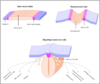
Formation of the neural tube

A

Neural folds become apposed and fuse. Some cells from the apices form the neural folds become separated to form the groups lying dorsolateral to the neural tube. These are known as neural crests.

The formation of neural tube is complete by the middle of the fourth week of embryonic development.

10
Q

Fate of neural crest cells

A

Form the sensory ganglia of the spinal and cranial nerves and also the autonomic ganglia
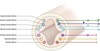
11
Q

Sulcus limitans

A

Longitudinal groove which appears on the inner surface of the lateral walls of the embronyic spinal cord and caudal part of the brain.

The dorsal and ventral cell groupings are thus delinated as the alar plate and basal plate

12
Q

Alar plate->

A

Predominantly sensory

13
Q

Basal plate

A

Predominantly motor

14
Q

Arrangement of neuronal cell groups in alar plate from ventral to dorsal

A

General visceral afferent

Special visceral afferent

General somatic afferent

Special somatic afferent

15
Q

Arrangement of neuronal cell groups (efferent) in the basal plate from ventral to dorsal

A

Somatic efferent

Branchial efferent

General visceral efferent

16
Q

Special somatic afferent

A

Associated with developing inner ear and ultimately receing auditory and vestibular input

17
Q

General somatic afferent

A

Receiving general sensory input from the periphery

18
Q

Special visceral afferent

A

Subserving the sense of taste

19
Q

General visceral afferent

A

Receiving afferent input from the viscera

20
Q

General visceral efferent

A

Preganglionic autonomic efferents

21
Q

Branchial efferent

A

Containing motor neurones to muscles derived from branchial arches

22
Q

Somatic efferent

A

Motor neurones to somatic muscles

23
Q

What are the three primary brain vesicles?

A

Prosencephalon

Mesencephalon

Rhombencephalon

24
Q

What are the two flexures of the developing CNS

A

Cephalix flexure

Cervical flexure

25
Site of the cephalic flexure
Midbrain forebrain
26
Location of the cervical flexure
Between the brain and the spinal cord
27
When are the three primary brain vesicles first seen
5th weeek
28
What are the five secondary brain vesicles
Telencephalon Diencephalon Mesencephalon Metencephalon Myelencephalon
29
Pontine flexure
Between metencephlon and myelencephalon
30
Telencephalon becomes
Cerebral hemisphere
31
Diencephalon becomes
Thalamus and related structures
32
Mesencephalon becomes
Midbrain
33
Metencephalon becomes
Pons, cerebellum
34
Myelencephalon becomes
Medulla oblongata
35
Corpora quadrigemina aka
Superior and inferior colliculi
36
Which sinus is contained in the tentorium cerebelli
Straight sinus
37
How many spinal nerves are there?
31
38
What happens to spinal nerves near to the cord
Divide into dorsal (posterior) and ventral (anterior) roots which attach to the cord along its dorslateral and ventrolateral bodies respectively.
39
Where are the cell bodies of the dorsal afferent fibres?
DRG
40
Where are the cell bodies of the ventral efferent neurones
In the spinal grey matter
41
Where does the spinal cord terminate in adults
L1 L2
42
Arrangement of grey matter in the spinal cord
Four extensions of central grey matter project dorsolaterally and ventrolaterally Dorsal and ventral horns.
43
Dorsal horns
Site of termination of many afferent neurones conveying impulses from the sensory receptors and is the site of origin of ascending pathways carrying sensory impulses to the brain
44
Ventral horn
Contains motor neurones that innervate skeletal muscles
45
Lateral horn of the spinal cord
Thoracic and upper lumbar levels of the SC have another smaller collection of cell bodies which contain preganglionic neurones belonging to the sympathetic division of the ANS
46
Which are the principle ascending tracts of the spinal cord?
Dorsal columns Spinothlamic Spinocerebellar tracts.
47
What are the four main divsions in a dorsoventral direction of the diencephalon
Epithalamus, thalamus, subthalamus, hypothalamus.
48
Epithalamus
Small and its most notable component is the pineal gland which lies in the midline, immediately rostral to the superior colliculi of the midbrain.
49
What arises from the ventral aspect of the hypothalamus in the midline?
Infundibulum/pituitary stalk which is attached to the pituitary galnd
50
51
52
Location of visual cortex
Occipital lobe mostly on the medial aspect of the hemisphere in the gyri above and below the calcarine sulcus
53
Corona radiata
Characteristic pattern of afferent and efferent connections between cortical and subcortical structures
54
What is the stereotyped arrangement of neurones carrying sensory information
First order Second order Third order
55
Passage of first order sensory neurones
Enter the spinal cord through a nerve or brainstem ipsilateral to the receptor. Cell body located in the DRG or trigeminal ganglion. Synapse ipsilaterally in the SC.
56
Passage of second order sensory neurone
Cell body in the spinal cord or brain stem. The exact location depends on the modality. Its axon decussates and ascends to the thalamus where it terminates
57
Passage of third order sensory neurone
Cell body in the thalamus, axon projects to the somatosensory cortex located in the postcentral gyrus of the parietal lobe of the cerebral hemisphere.
58
Where do axons carrying information from the spinothalamic tract decussate.
Second order neurones carrying information from the spinothalamic tract decussate at the level of the SC
59
Where do first order sensory neurones carrying dorsal columnar information terminate?
Synapse with dorsal column nuceli (nuceli gracilis and cuneatus) in the medulla. The second order sensory neurones decussate and ascend to the thalamus
60
In what tract do second order sensory neurones carry dorsal column information to the thalamus after decussation at the level of the medulla?
The medial lemniscus
61
Passage of primary afferent neurones of the trigeminal nerve
Enter the brainstem and terminate ipsilaterally in the trigeminal sensory nucleus. Second order neurones decussate and ascend to the thalamus as the trigeminothalamic tract.
62
On what portion of the thalamus do second order sensory neurones converge?
The ventral posterior nucleus of the thalmus.
63
What are the two main descending tracts?
Corticospinal Corticobulbar
64
Descending pathways
UMN-\> LMN which has cell body in the grey matter of the SC and brain stem.
65
What proportion of UMNs decussate at the pyramidal decussation?
90%, 10% continue on the same side as the anterior corticospinal tract
66
Name of the location of UMN decussation?
Decussation of the pyramids.
67
What are the extrapyramidal descending tracts
Vestibulospinal Reticulospinal
68
How can the four major disorder types of neuromuscular system be classified?
Extrinsic Systemic Vacular Intrinsic
69
Extrinsic neuromuscular pathology
Lead to compression of brain SC nerve roots and peripheral nereves. Are thus surgically remediable.
70
Syrinx
Expansion of the minute vestigial space of the SC into a cavity.
71
Dissociated sensory loss
Clinical finding of selective loss of the modalities of touch/proprioceptino with preservation of pain and temperature or vice versea
72
Pyramidal weakness
Weakness of extension and abduction in upper limb Weakness of flexion in lower limb
73
Romberg's sign
Indicative of sensory ataxia rather than lesions of cerebellar pathways
74
Agnosia
Loss of perception
75
Apraxia
Loss of skilled movements
76
During development the brain can be divided into five contiguous parts. Which part of the embryonic brain gives rise to the cerebellum? Metencephalon Telencephalon Diencephalon Myelencephalon Mesencephalon
Metencephlaon