Spinal Neurosurgery Flashcards
(86 cards)
A 37-year-old man, normally active and otherwise healthy, develops a sudden pain in his back and an ‘electric shock’ down his left leg. Over 5 weeks the back pain resolves but he is left with intermittent severe pain in the left leg which radiates down his calf into the sole of the foot and little toe. His right leg has been unaffected and he has normal sphincter function.
What is the likely diagnosis? Is there a differential diagnosis?
The history is classical for a prolapsed L5/S1 intervertebral disc and left S1 dermatomalsciatica (see ‘ Nerve roots affected by a prolapsed disc ’, p. 328).
Hip and knee pathology can both mimic sciatica, especially in older people. Many patients will have back pain, and there may be dual pathology. Recent exacerbation of back pain with sciatica suggests the two are linked. Rarely, acute leg pain can be a presentation of vascular events such as limb ischaemia. Calf pain may be a symptom of DVT which should be considered in a patient with severe immobility due to back pain
A 37-year-old man, normally active and otherwise healthy, develops a sudden pain in his back and an ‘electric shock’ down his left leg. Over 5 weeks the back pain resolves but he is left with intermittent severe pain in the left leg which radiates down his calf into the sole of the foot and little toe. His right leg has been unaffected and he has normal sphincter function.
The patient walks with a mildly antalgic gait. What other signs would you look for on examination?
Most common finding will be restriction of passive straight leg raise
May have senosry change in the left S1 dermatome and absent or diminished ankle jerk.
In more severe cases there is S1 numbness and weakness of ankle plantarflexion.
He has restricted straight leg raise at 40 degrees, despite mild senosry loss and weak ankle jerk, he is sensorily intact.
What is the diagnosis and how would you manage him at this stage?
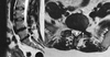
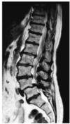
There are two degenerate discs at L4/5 (A) and L5/S1 (B)
The L5/S1 bulges posteriorly a bit more than the L4/5 and the axial slices show a left posterolateral disc prolapse compressing the left S1 nerve root (C). The left S1 root is not visible separate from the disc.
Most disc prolapses settle with conesrvative management- rest and analgesia int he first instance.
If there is a neurological deficit there is a stronger case for early surgery, especially if foot drop is present.
The point at which surgery is considered depends on degree of pain and impact on QoL
6/52 typically is what is trialled for conservative Mx
37-year-old man, normally active and otherwise healthy, develops a sudden pain in his back and an ‘electric shock’ down his left leg. Over 5 weeks the back pain resolves but he is left with intermittent severe pain in the left leg which radiates down his calf into the sole of the foot and little toe. His right leg has been unaffected and he has normal sphincter function.
What surgery would you offer?
As leg pain is the main complaint- lumbar discectomy for the L5/S1 fragment.
Discectomy for leg pain with typical symptoms and clinical-radiological correlation has a high chance (>90% of improving his leg pain)
There may be secondary improvement in his back pain
Risks of lumbar discectomy
Haemorrhage
Infection
Infective discitis- results in progressive back pain and rarely, sepsis in the weeks following surgery which would require prolonged Abx
The risk of neurological complications e.g. cauda equina should be small.
Risk of S1 weakness should be mentioned as a rare occurence.
Early recurrence in the first few weeks following surgery can be treated with repeat surgery with good outcome
A 38-year-old woman presents to the emergency department with 2 weeks of back pain radiating down her left leg. On the previous day, she noticed difficulty passing urine and partial numbness of her buttocks.
What is the diagnosis?
Cauda equina until proven otherwise
Def: Cauda equina syndrome
Clinical syndrome resulting from the compression of the cauda equina.
Presents with a variable combination of:
leg pain (classically bilateral)
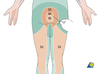
sacral anaesthesia
urinary retention with overflow incontinence.
Causes of cauda equina compression
Prolapsed lumbar intervertebral disc.
Tumours, abescess, trauma can also cause the syndrome
Important questions in cauda equina?
Characterise the exact nature of urinary symptoms as urinary retention can occur due to back pain.
Timing of onset of symptoms is also critical as it informs the timing of surgery.
Patient with incomplete cauda equina syndrome have better prognosis
A 38-year-old woman presents to the emergency department with 2 weeks of back pain radiating down her left leg. On the previous day, she noticed difficulty passing urine and partial numbness of her buttocks.
On examination there was some weakness of plantarflexion and
hip extension on the left. The left ankle reflex was absent.
Pinprick sensation was preserved throughout the lower limbs
but was absent in the perineum. However, when more pressure
was applied to the pin, the patient reported that she could feel
the sharp pin in the perineum. The rest of the examination
was normal.
Level of lesion
Left ankle is affected suggesting S1 involvement
Ankle plantar flexion and hip extension also have contributions from S1.
Level of compression is likely to be at L5/S1
A 38-year-old woman presents to the emergency department with 2 weeks of back pain radiating down her left leg. On the previous day, she noticed difficulty passing urine and partial numbness of her buttocks.
On examination there was some weakness of plantarflexion and
hip extension on the left. The left ankle reflex was absent.
Pinprick sensation was preserved throughout the lower limbs
but was absent in the perineum. However, when more pressure
was applied to the pin, the patient reported that she could feel
the sharp pin in the perineum. The rest of the examination
was normal.
Are the clinical findings consistent with the suspected diagnosis?
Examining perineal sensation can be challenging if the patient’s response is inconsistent. As a general rule, if perineal pinprick sensation is less than reported elsewhere when the same pressure is applied, it should be assumed that there is impairment. If a sharp sensation is elicited when the pin is pressed harder, impairment is not excluded. If a sharp sensation is not elicited at all, impairment is confirmed. Therefore these findings are consistent with cauda equina compression.
Ix in ?cauda equina
MRI lumbar spine
other options include CT myelogram or MRI under GA
?Dx

Large disc prolapse at L5/S1 filling the width of the canal (1).
Axial view shows the disc is laterally sited to the left. (2)
The thecal sac which appears white on the T2 image is displaced to the right.
There is a samlller left sided protrusion at the level above where the canal is more capacious,.
Mx of cauda equina
Discectomy involving mid-line posterior approch, laminectomy and removal of the prolapse segment of the disc.
A 38-year-old woman presents to the emergency department with 2 weeks of back pain radiating down her left leg. On the previous day, she noticed difficulty passing urine and partial numbness of her buttocks.
She undergoes surgery to decompress the cauda equina.
The patient is reviewed the following morning when
she complains of recurrent left leg pain. How should
her symptoms be managed?
DDx:
Residual disc
Post-operative haematoma
Infection
Post-surgical oedema.
Repeat MRI is required to exclude persistent neural compression
In the presence of what particular feature should you have a low threshold for emergency scanning in ?cauda equina?
Sensory changes in the sacral deramtomes, S2, 3 or 4 even if only subjective or unilateral.
Relationship between nerve root and intervertebral dsic
Situated just above the level of the numbered disc.
Unilateral lumar disc prolapse tends to ocompression the transversing nerve roots whereas a far lateral prolapse will compress the exiting nerve root at the levl.

A 43-year-old woman has a 1-year history of right arm pain felt around the elbow and radiating into the thumb and index finger. It has settled a little but she has a continu- ous tingling feeling in the same region. There is no history of neck pain or trauma.
Ddx and key features of Hx or exam
Neurological or MSK pain.
The tingling suggests the symptoms are more likely to be neurological in origin.
2 most likely are CTS which may radiate proximally beyond the wrist to the elbow or shoulder. And radiculopathy, classically C6 if the thumb and index finger are involved.
MSK causes such as lateral epicondylitis and tenosynovitis should be considered and can be excluded by point tenderness at the site of inflammation exacerbated by passive movement.
CTS usually repsonds to Tinel’s/Phalen’s test.
Cervical radicular pain can be reproduced by asking the patient to look up and turn their head to the contralateral side, to reduce the calibre of the affected nerve root foramen and reproduce symptoms
Spurling’s sign
Reproduction of radicular pain on looking up and turning head to contralateral side, which narrows the foramen.
A 43-year-old woman has a 1-year history of right arm pain felt around the elbow and radiating into the thumb and index finger. It has settled a little but she has a continu- ous tingling feeling in the same region. There is no history of neck pain or trauma.
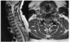
On closer assessment she confirms that the thumb and index finger, rather than the thumb and the lateral two and a half fingers, are affected. Spurling’s sign is positive and you arrange an MRI. The T2 sagittal images of the midline and to the right of the midline and the axial image of the C5/6 disc are shown in Fig. 48.1. Comment on the findings.

There is loss of cervical lordosis, which is very common in patients with acute and chronic cevical spine pathology.
The discs apppear healthy on the midline image and there is no loss of disc height or osteophyte formation.
The saggital image to the right of the midline shows some loss of CSF signal adjacent to the disc at the C5/6 level.
The axial scans of C5/6 show the left C6 nerve root with the CSF surrounding it, but there is narrowing of the right C6 root due to lateral disc or osteophyte
A 43-year-old woman has a 1-year history of right arm pain felt around the elbow and radiating into the thumb and index finger. It has settled a little but she has a continu- ous tingling feeling in the same region. There is no history of neck pain or trauma.
MRI shows narrowing of the C6 nerve root and the surrounding CSF due to osteophyte or canal narrowing.
What are the options?
She does not have a progressive deficit and surgery should be offered if pain is intractable or intolerable.
A radiologically guided nerve root infiltration with steroids may offer temproary relief.
Surgical options for C6 nerve root compression
Options are anterior cervical discectomy (ACD) or foraminotomy (posterior approach to de-roof the right C6 neural foramen) as the aim is to decompress the C6 nerve root at the C5/6 neural foramen
What are the benefits of ACD
Will remove primary pathology (disc prolapse) and has a higher success rate in alleviating the brachalgia (95%)
What are the possible complications of ACD
Significant potential morbidity from complications such as RLN palsy, oesophageal, carotid injury and neck wound haematomas