Histopathology 11 - Dermatopathology Flashcards
(45 cards)
Recall the 3 broad layers of the skin
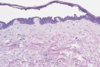
Epidermis, dermis and subcutaneous fat = ~6 mm thickness
Recall the layers of the epidermis
Layers of skin: “Come, let’s get some beers”

What structures are present in the dermis?
blood vessels, sweat glands, hair follicles, sebaceous glands and nerve fibres
Recall examples of vesiculobullous inflammation?
Bullous pemphigoid
Pemphigous vulgaris
Pemphigus foliaceus
What are the aetiological agents of pemphigoid
IgG and C3 attack basement membrane
Eosinophils recruited → elastase → damages anchoring proteins → fluid fills up gap between BM and epithelium

What is the presentation of bullous pemphigoid?
Elderly, autoimmune, high mortality
Flexor surfaces, tense bullae
Dermo-epidermal junction

What are the histological features of bullous pemphigoid
↑ eosinophils
How can you confirm the diagnosis of pemphigoid?
Immunofluorescence shows IgG along basement membrane
IgG anti-hemidesmosome

What causes pemphigus vulgaris?
Epiderma-epidermal junction affected
IgG attacks between keratin layers (acantholysis) → loss of intracellular connections
in stratum spinosum
What is the presentation of pemphigus vulgaris?
Flaccid blisters, rupture easily
What is pemphigus foliaceus?
Top layer very thin so never blisters
IgG-mediated – outer layer of stratum corneum shears off
Diagnose with immunofluorescence
Give examples of spongiotic skin inflammation
Discoid eczema
Contact dermatitis
On which surfaces does psoriasis tend to present?
Extensor with silver plaques

On which surfaces does eczema tend to present?
Flexor
What is the pathophysiology for spongiotic skin inflammation?
Itchy → hyperparakeratosis (thickening) → lichenification
Epidermis thicker
Eczema is spongiotic - oedema between keratinocytes
T cell mediated and eosinophils are recruited
Which cells of the immune system are most involved in eczema?
T-cell mediated pathology
Eosinophils recruited to sites of inflammation
Recall the pathophysiology of psoriasis
Normal keratinocyte turnover time = 56 days
Psoriasis keratinocyte turnover time = 7 days
- Rapid turnover → epidermis thicker
- layer of parakeratosis at the top
- Stratum granulosum disappears as not enough time to form it; and dilated vessels form
- Munro’s microabscesses form, made up from recruitment of neutrophils
Which immune-mediated skin condition causes a rapid turnover of keratinocytes?
Psoriasis
Recall the pathophysiology for lichen planus
T-cell mediated; itchy
T-lymphocytes destroy bottom keratinocytes
Creates band-like inflammation
Cannot see where dermis finished, and epidermis starts
How does lichen planus present?
Papules and plaques of purplish-red colour on wrists and arms
In mouth it presents as white lines (Wickham striae)
Which skin condition appears as white lines?
Lichen planus
Which skin pathology appears as “silvery plaques”?
Psoriasis
Where does fluid build in eczema?
Between keratinocytes
What is pyoderma gangrenosum and what might it be a sign of?
non-healing ulcer
Often, first manifestation of a systemic disease - colitis, hepatitis, leukaemia