Histopathology 2 - Bone tumours Flashcards
(35 cards)
What is the preferred investigation for diagnosing bone tumours?
US-guided Jamshidi needle biopsy
Recall 4 tumour-like conditions of the bone that are not actually malignant
- Fibrous dysplasia
- Fibroma (can be ossifying/ non-ossifying)
- Reparative giant cell granuloma
- Simple bone cyst
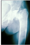
What is “shepherd’s crook deformity” a reference to?
Fibrous dysplasia involving the femoral head
(ribs and proximal femur most common)
Microfractures → change in shape

Which syndrome is fibrous dysplasia associated with ?
McCune Albright Syndrome
polyostotic fibrous dysplasia + rough border café au lait spots on the skin
Which mutation is involved in fibrous dysplasia?
Caused by a mutation in a G-protein (GNAS mutation chr20; q13)
What XR finding would be present in fibrous dysplasia?
‘soap bubble’ osteolysis
What histological features would be present in fibrous dysplasia?
marrow replaced by fibrous stroma with rounded trabecular bone (used to be called Chinese letters)

How does osteochondroma mimic bone in appearance?
cartilaginous surface overlying normal cortical + trabecular bone
at ends of long bones
What is the typical age of presentation of osteochondroma?
male 10-20y
Where is the cartilaginous proliferation in enchodroma?
Cartilaginous proliferation within the bone
hands and feet (rarely at the end of long bones)
How will enchondroma appear on XR?
“popcorn” pattern
In which bones is osteochondroma most likely to present?
Long bones
What are some benign bone forming tumours?
Osteoid osteoma
Osteoblastoma
Osteoma
Is a giant cell tumour of bone benign or malignant?
Borderline malignant
What is the X ray appearance of giant cell tumour?
lytic appearance
What is the typical age of presentation of giant cell tumour of bone?
20-40 years females
Female version of osteochondroma
How do giant cell bone tumours appear under the microscope?
Osteoclasts on background of spindle/ovoid cells
‘Giant Cell’ - composed mostly of osteoclast giant cells, but NOT the neoplastic cells (tumour cells = stromal cells)
What is the most common malignant bone tumour?
MOST COMMON malignant bone tumour (mets v rare below elbow and knee)
What are the 3 types of malignant bone tumour?
Osteosarcoma (bone-forming)
Chondrosarcoma (cartilage-forming)
Ewing’s sarcoma (undifferentiated mesenchymal)
Recall the typical age of presentation for each of the 3 types of malignant bone tumour
Osteosarcoma: <30 years, MALES
Chondrosarcoma: >40 years
Ewing’s sarcoma: <25 years
Recall the typical site affected for each of the 3 types of malignant bone tumour
Osteosarcoma: knee (end of long bones)
Chondrosarcoma: axial, pelvis/ proximal skeleton
Ewing’s sarcoma: diaphysis/metaphysis of long bones, pelvis
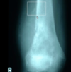
Recall the typical X ray appearance of each of the 3 types of malignant bone tumour
Osteosarcoma: Codman’s triangle
Chondrosarcoma: fluffy calcification
Ewing’s sarcoma: Onion-skinning of periosteum
What is a “Codman’s triangle”?
The triangular area of new subperiosteal bone that is created when a lesion, often a tumour, raises the periosteum away from the bone.
Which of the 3 types of malignant bone tumour has the best prognosis?
Chondrosarcoma
