Histopathology 15 - Cerebrovascular disease and Trauma Flashcards
(38 cards)
What are the 2 main types of cerebral oedema?
Vasogenic (due to disrupted BBB)
Cytotoxic (due to cellular injury e.g. hypoxia/ ischaemia)
where does cytotoxic cerebral oedema affect?
damage at astrocyte end-foot processes
What water channel is used to transport water molecules in the brain?
AQA4
What is the characteristic radiological finding in cerebral oedema?
loss of gyri

Describe the normal CSF flow in the brain
CSF made in choroid plexus (mainly in lateral ventricles) → lateral ventricles → 3rd ventricle → cerebral aqueduct → 4th ventricle
CSF flows down → medulla → central canal of the spinal cord
Relatively little CSF volume will go down spinal cord - most exits via foramina in 4th ventricle → subarachnoid space
CSF → subarachnoid space and via arachnoid granulations which pierce superior sagittal sinus, thereby returning CSF to systemic circulation
What is the difference between communicating and non-communicating hydrocephalus?
Communicating = problem with CSF resorption into venous sinuses (no obstruction) - infection/inflammation
Non-communicating = obstruction to CSF flow (usually cerebral aqueduct)
What is the normal range for ICP in a supine adult?
7-15mmHg
What is the most important contraindication to lumbar puncture?
Pailloedema
What is the most common site for non-traumatic intra-parenchymal haemorrhages?
Basal ganglia
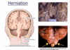
what is a complication of ↑ICP HERNIATION of brain structures where space is available?
HERNIATION of brain structures where space is available
What are the THREE sites of brain herniation?
- Subfalcine - cortex forced under rigid falx cerebri
- Uncal (transtentorial) - medial temporal lobe through tentorial notch
- Tonsillar - tonsil of cerebellum pushed through foramen magnum
Define stroke
rapidly developing clinical symptoms and/or signs of focal, and at times global loss of cerebral function, with symptoms > 24 hours or leading to death, with no apparent cause other than that of vascular origin
What is the main cause of infarct and where is it most commonly seen?
mostly from cerebral atherosclerosis
Particularly bad at carotid bifurcation or basilar artery
also from emboli from heart (i.e. AF) → MCA branches
focal vs global cerebral ischaemia
Focal cerebral ischaemia – due to lack of blood flow to a particular vascular territory
Global cerebral ischaemia – when the systemic circulation fails
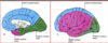
MCA vs ACA supply territories
MCA supplies the OUTSIDE, ACA supplies the middle & front
What is the most common type fo haemorrhagic stroke?
sub-arachnoid (most common) haemorrhage
also primary intracerebral, intraventricular
What is the biggest risk factor for non-traumatic intra-parenchymal haemorrhage?
Hypertension (>50% of bleeds)
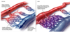
When do congenital arteriovenous malformations tend to become symptomatic?
Between 2nd and 5th decade
How doe AV malformations cause a stroke?
high pressure → MASSIVE BLEED
What is the management of ruptured congenital arteriovenous malformation?
Surgically remove if poss, this may be radiosurgery
Embolise (to stop bleeding)
What is a cavernous angioma?
tightly packed vessels - no brain parenchyma in between vascular spaces” → similar to an AVM but no brain substance wrapped up amongst vessels
When do cavernous angiomas become symptomatic?
When they bleed - which is at low pressure and usually >50 years
What is the most common site of haemorrhage in subarachnoid haemorrhage?
Berry aneurysm rupture
80% at internal carotid artery bifurcation
What characteristic sign can you visualise on MRI of cavernous angioma?
T2-weighted “Target Sign” – black ring around lesion (AVM has no ring) – no brain parenchyma


